
Cryptosporidium
Synopsis
CAPC Recommends
- Continuing to monitor the health of the pet for those dogs that test positive for Cryptosporidium spp. on annual wellness fecal testing.
- Diagnosing and treating any underlying conditions that may be weakening the animal’s immune system or causing stress, which can allow Cryptosporidium species to multiply.
- Using diet and supplements to support GI health and normalize the GI biome.
Introduction
- Cryptosporidium species are coccidian-like protozoan parasites that infect a wide variety of vertebrate hosts.
- Most species and strains are highly adapted to specific hosts.
- When other hosts are exposed to these strains, they are less likely to be infected unless they are immunocompromised.
- Humans are more likely to become infected by C. hominis and certain genotypes of C. parvum than by either a canine or feline species.
- However, some species are thought to be shared among dogs, cats, and humans.
Stages
- Sporulated oocysts—resistant stages for environmental transmission—are infective when shed by a host.
Schizonts (asexual stages)
Gametes (sexual stages)

Oocysts of Cryptosporidium from fecal flotation

Photomicrograph of developmental stages of Cryptosporidium from the small intestine

Scanning electron microscopy showing Cryptosporidium stages in the microvillus border of epithelial cells
Disease
- Cryptosporidiosis is a self-limited, small bowel infection characterized by secretory diarrhea. Fluid loss may be severe in clinical cases.
- The disease may be more severe, prolonged, and sometimes life-threatening in immunocompromised hosts.
- In many cases dogs may be asymptomatic.
Prevalence
Seroprevalence along with microscopic identification and PCR-based diagnostic studies indicate that Cryptosporidium infection is relatively common in dogs.
Confirmation of current infection by fecal examination is infrequent; commonly seen in only 1% to 5% of cases. Seroprevalence (antibody) studies, on the other hand, suggest higher rates of resolved infection; antibody titers are measurable in 20% to 50% of cases.
Transmission
- Infection occurs following ingestion of sporulated oocysts from fecal-contaminated environments, food, or water.
Host Associations and Transmission Between Hosts
- Cryptosporidium exists as several distinct species and genotypes with different degrees of infectivity for animals and humans:
- C. canis - dogs, rarely humans
- C. felis - cats, rarely humans
- C. hominis - only in humans
- C. parvum - a species in ruminants and other hosts that also will readily infect people.
- Although Cryptosporidium canis and Cryptosporidium felis primarily infect dogs and cats, respectively, zoonotic transmission to humans has been documented. See section on 'Zoonotic Potential of Valid Species and Genotypes of Cryptosporidium' for more information.
Prepatent Period and Environmental Factors
- Oocysts are generally shed in the feces 3 to 6 days after infection.
Oocysts are immediately infectious when passed and are capable of surviving in the environment for extended periods.
Oocysts are resistant to most disinfectants, including routine chlorine concentrations in drinking water and swimming pools.
Oocysts are susceptible to commercial formulations of ammonia and heat over 70°C.
Site of Infection and Pathogenesis
- Cryptosporidium spp. infect the microvillar border of cells lining the small intestine and other organs. The developmental cycle is similar to Cystoisospora spp.
- In immmunocompromised humans, infections may also occur in the large intestine, respiratory tract, biliary tract, pancreatic duct, and other sites.
- Most Cryptosporidium infections in dogs are subclinical.
- Diarrhea and dehydration are the primary clinical signs
- In immunocompetent and healthy dogs, the infections are usually self-limiting and some may be asymptomatic.
- Persistent infections usually denote an underlying cause.
Diagnosis
- Diagnosis can be difficult. Specimens should be sent to an academic or commercial testing laboratory familiar with the necessary diagnostic techniques.
- Specific species identification requires molecular methods (e.g., PCR) that are currently only performed by some reference laboratories as well as several academic laboratories.
- Oocysts are typically isolated using centrifugal flotation with sucrose.
- The oocysts are spheroid and small, 4-6 µm in diameter. In sucrose preparations, the oocysts float just under the coverslip on the slide.
- Oocysts are clear or the walls may have pinkish hue depending on the objective lens used.
- These are some of the smallest parasites seen in fecal samples and require skill and practice for accurate diagnosis.
- Oocysts in acid fast stained fecal smears are red or pink.
- Fluorescent antibody tests (IFA) can be performed on fecal smears.
- Fecal antigen is detected with enzyme-linked immunosorbent assays (ELISAs).
- Fecal PCR tests for Cryptosporidium
- Commercial assays are available for detection of Cryptosporidium DNA.
- Nucleic acid is stable in the feces for 10 days at 4ºC.
- Both centrifugal fecal flotation and fecal PCR have their strengths and weaknesses, however to ensure the widest breadth of detection of intestinal parasites in dogs, fecal PCR tests should be combined with microscopic examination of feces.
- Commercial laboratories publish algorithms specific to their diagnostic tests and may be useful.
- Patient status and lifestyle are important considerations when interpreting Cryptosporidium positive diagnostic results.
Treatment
- Treatment is difficult and few drugs are consistently effective against Cryptosporidium.
- Symptomatic patients may require treatment, particularly if clinical signs are severe or persistent.
- Non-symptomatic patients may not require treatment.
- Patients living in a household with vulnerable or immunocompromised humans or pets should be treated.
- Ultimately, it is the practitioner's responsibility to use their best judgement in consultation with the owner in deciding treatment options.
- The following drugs have been used with some success in cases where animals have persistent diarrhea with oocyst shedding:
- Azithromycin: 5 to 10 mg/kg BID for 5 to 7 days (dogs)
- Until recently nitazoxanide was approved only for use in humans. While there are few studies in dogs, and none in cats, one study found that treatment with either 75 mg/kg or 150 mg/kg was effective in clearing Cryptosporidium spp. in dogs compared to controls.
- Diagnosing and treating any underlying immunosuppressive conditions is key to managing Cryptosporidium app. in those patients that are ill.
- A key component to treatment may be normalizing the GI biome, which can be aided by probiotics and/or diets specifically formulated to support GI health.
Control and Prevention
- Due to the low infective dose and persistence of oocysts in the environment coupled with the lack of effective treatment, control and prevention should be focused on reducing environmental contamination with infectious oocyst and should consist of:
- Promptly picking up and removing feces to prevent fecal contamination of the environment.
- Cleaning with heat or steam followed by disinfection and drying to promote desiccation, may be useful.
Public Health Considerations
- The zoonotic risk of all recognized Cryptosporidium species and genotypes is not known. However, 19 species and 4 genotypes have been identified in humans (See “Zoonotic Potential of Valid Species and Genotypes of Cryptosporidium” for more information).
- Human infections are most often due to C. hominis and C. parvum.
- Other species more commonly implicated in human infections are C. meleagridis, C. felis, C. canis, and C. ubiquitum. However, the risk for acquiring these Cryptosporidium species in comparison to C. hominis and C. parvum is much lower.
- In the United States, Cryptosporidium infections are the leading cause of waterborne illness in humans.
- To reduce the risk of acquiring Cryptosporidium infections, people should practice good hygiene, avoid swimming in potentially contaminated water, and avoid drinking untreated water or consuming potentially contaminated food.
Zoonotic Potential of Valid Species and Genotypes of Cryptosporidium
- There are 44 recognized species and numerous genotypes of Cryptosporidium.
- Of these, 19 species and 4 genotypes have been identified in human infections either from immunocompetent or immunocompromised individuals. Below is a summary of the Cryptosporidium species and genotypes found in human infections as well as their primary host species.
C. hominis
- Primary Host: Humans
C. parvum
- Primary Host: Many mammals
C. meleagridis
- Priary Host: Avians
C. canis
- Primary Host: Dogs
C. felis
- Primary Host: Cats
C. ubiquitum
- Primary Host: Many mammals
C. cuniculus
- Primary Host: Rabbits
C. viatorum
- Primary Host: Rodents
C. muris
- Primary Host: Rodents
C. andersoni
- Primary Host: Cattle
C. erinacei
- Primary Host: Hedgehogs
C. tyzzeri
- Primary Host: Rodents
C. bovis
- Primary Host: Cattle
C. suis
- Primary Host: Pigs
C. scrofarum
- Primary Host: Pigs
C. occultus
- Primary Host: Rodents
C. xiaoi
- Primary Host: Small ruminants
C. fayeri
- Primary Host: Marsupials
C. ditrichi
- Primary Host: Rodents
Chipmunk genotype I
- Primary Host: Rodents
Mink genotype
- Primary Host: Mink
Skunk genotype
- Primary Host: Skunk
Horse genotype
- Primary Host: Horse
Selected References
- Barbosa AD, Egan S, Feng Y, Xiao L, Ryan U. 2023. Cryptosporidium and Giardia in cats and dogs: What is the real zoonotic risk? Curr Res Parasitol Vector Borne Dis 4: Article 100158.
- Lucio-Forster A, et al, 2010. Minimal zoonotic risk of cryptosporidiosis from pet dogs and cats. Trends Parasitol., 6:4:174.
- Ryan U, Zahedi A, Feng Y, Xiao L. 2021. An update on zoonotic Cryptosporidium species and genotypes in humans. Animals (Basel) 11: Article 3307.
Synopsis
CAPC Recommends
- Continuing to monitor the health of the pet for those dogs that test positive for Cryptosporidium spp. on annual wellness fecal testing.
- Diagnosing and treating any underlying conditions that may be weakening the animal’s immune system or causing stress, which can allow Cryptosporidium species to multiply.
- Using diet and supplements to support GI health and normalize the GI biome.
Introduction
- Cryptosporidium species are coccidian-like protozoan parasites that infect a wide variety of vertebrate hosts.
- Most species and strains are highly adapted to specific hosts.
- When other hosts are exposed to these strains, they are less likely to be infected unless they are immunocompromised.
- Humans are more likely to become infected by C. hominis and certain genotypes of C. parvum than by either a canine or feline species.
- However, some species are thought to be shared among dogs, cats, and humans.
Stages
- Sporulated oocysts—resistant stages for environmental transmission—are infective when shed by a host.
Schizonts (asexual stages)
Gametes (sexual stages)
Oocysts of Cryptosporidium from fecal flotation
Photomicrograph of developmental stages of Cryptosporidium from the small intestine
Scanning electron microscopy showing Cryptosporidium stages in the microvillus border of epithelial cells
Disease
- Cryptosporidiosis is a self-limited, small bowel infection characterized by secretory diarrhea. Fluid loss may be severe in clinical cases.
- The disease may be more severe, prolonged, and sometimes life-threatening in immunocompromised hosts.
- In many cases dogs may be asymptomatic.
Prevalence
Seroprevalence along with microscopic identification and PCR-based diagnostic studies indicate that Cryptosporidium infection is relatively common in dogs.
Confirmation of current infection by fecal examination is infrequent; commonly seen in only 1% to 5% of cases. Seroprevalence (antibody) studies, on the other hand, suggest higher rates of resolved infection; antibody titers are measurable in 20% to 50% of cases.
Transmission
- Infection occurs following ingestion of sporulated oocysts from fecal-contaminated environments, food, or water.
Host Associations and Transmission Between Hosts
- Cryptosporidium exists as several distinct species and genotypes with different degrees of infectivity for animals and humans:
- C. canis - dogs, rarely humans
- C. felis - cats, rarely humans
- C. hominis - only in humans
- C. parvum - a species in ruminants and other hosts that also will readily infect people.
- Although Cryptosporidium canis and Cryptosporidium felis primarily infect dogs and cats, respectively, zoonotic transmission to humans has been documented. See section on 'Zoonotic Potential of Valid Species and Genotypes of Cryptosporidium' for more information.
Prepatent Period and Environmental Factors
- Oocysts are generally shed in the feces 3 to 6 days after infection.
Oocysts are immediately infectious when passed and are capable of surviving in the environment for extended periods.
Oocysts are resistant to most disinfectants, including routine chlorine concentrations in drinking water and swimming pools.
Oocysts are susceptible to commercial formulations of ammonia and heat over 70°C.
Site of Infection and Pathogenesis
- Cryptosporidium spp. infect the microvillar border of cells lining the small intestine and other organs. The developmental cycle is similar to Cystoisospora spp.
- In immmunocompromised humans, infections may also occur in the large intestine, respiratory tract, biliary tract, pancreatic duct, and other sites.
- Most Cryptosporidium infections in dogs are subclinical.
- Diarrhea and dehydration are the primary clinical signs
- In immunocompetent and healthy dogs, the infections are usually self-limiting and some may be asymptomatic.
- Persistent infections usually denote an underlying cause.
Diagnosis
- Diagnosis can be difficult. Specimens should be sent to an academic or commercial testing laboratory familiar with the necessary diagnostic techniques.
- Specific species identification requires molecular methods (e.g., PCR) that are currently only performed by some reference laboratories as well as several academic laboratories.
- Oocysts are typically isolated using centrifugal flotation with sucrose.
- The oocysts are spheroid and small, 4-6 µm in diameter. In sucrose preparations, the oocysts float just under the coverslip on the slide.
- Oocysts are clear or the walls may have pinkish hue depending on the objective lens used.
- These are some of the smallest parasites seen in fecal samples and require skill and practice for accurate diagnosis.
- Oocysts in acid fast stained fecal smears are red or pink.
- Fluorescent antibody tests (IFA) can be performed on fecal smears.
- Fecal antigen is detected with enzyme-linked immunosorbent assays (ELISAs).
- Fecal PCR tests for Cryptosporidium
- Commercial assays are available for detection of Cryptosporidium DNA.
- Nucleic acid is stable in the feces for 10 days at 4ºC.
- Both centrifugal fecal flotation and fecal PCR have their strengths and weaknesses, however to ensure the widest breadth of detection of intestinal parasites in dogs, fecal PCR tests should be combined with microscopic examination of feces.
- Commercial laboratories publish algorithms specific to their diagnostic tests and may be useful.
- Patient status and lifestyle are important considerations when interpreting Cryptosporidium positive diagnostic results.
Treatment
- Treatment is difficult and few drugs are consistently effective against Cryptosporidium.
- Symptomatic patients may require treatment, particularly if clinical signs are severe or persistent.
- Non-symptomatic patients may not require treatment.
- Patients living in a household with vulnerable or immunocompromised humans or pets should be treated.
- Ultimately, it is the practitioner's responsibility to use their best judgement in consultation with the owner in deciding treatment options.
- The following drugs have been used with some success in cases where animals have persistent diarrhea with oocyst shedding:
- Azithromycin: 5 to 10 mg/kg BID for 5 to 7 days (dogs)
- Until recently nitazoxanide was approved only for use in humans. While there are few studies in dogs, and none in cats, one study found that treatment with either 75 mg/kg or 150 mg/kg was effective in clearing Cryptosporidium spp. in dogs compared to controls.
- Diagnosing and treating any underlying immunosuppressive conditions is key to managing Cryptosporidium app. in those patients that are ill.
- A key component to treatment may be normalizing the GI biome, which can be aided by probiotics and/or diets specifically formulated to support GI health.
Control and Prevention
- Due to the low infective dose and persistence of oocysts in the environment coupled with the lack of effective treatment, control and prevention should be focused on reducing environmental contamination with infectious oocyst and should consist of:
- Promptly picking up and removing feces to prevent fecal contamination of the environment.
- Cleaning with heat or steam followed by disinfection and drying to promote desiccation, may be useful.
Public Health Considerations
- The zoonotic risk of all recognized Cryptosporidium species and genotypes is not known. However, 19 species and 4 genotypes have been identified in humans (See “Zoonotic Potential of Valid Species and Genotypes of Cryptosporidium” for more information).
- Human infections are most often due to C. hominis and C. parvum.
- Other species more commonly implicated in human infections are C. meleagridis, C. felis, C. canis, and C. ubiquitum. However, the risk for acquiring these Cryptosporidium species in comparison to C. hominis and C. parvum is much lower.
- In the United States, Cryptosporidium infections are the leading cause of waterborne illness in humans.
- To reduce the risk of acquiring Cryptosporidium infections, people should practice good hygiene, avoid swimming in potentially contaminated water, and avoid drinking untreated water or consuming potentially contaminated food.
Zoonotic Potential of Valid Species and Genotypes of Cryptosporidium
- There are 44 recognized species and numerous genotypes of Cryptosporidium.
- Of these, 19 species and 4 genotypes have been identified in human infections either from immunocompetent or immunocompromised individuals. Below is a summary of the Cryptosporidium species and genotypes found in human infections as well as their primary host species.
C. hominis
- Primary Host: Humans
C. parvum
- Primary Host: Many mammals
C. meleagridis
- Priary Host: Avians
C. canis
- Primary Host: Dogs
C. felis
- Primary Host: Cats
C. ubiquitum
- Primary Host: Many mammals
C. cuniculus
- Primary Host: Rabbits
C. viatorum
- Primary Host: Rodents
C. muris
- Primary Host: Rodents
C. andersoni
- Primary Host: Cattle
C. erinacei
- Primary Host: Hedgehogs
C. tyzzeri
- Primary Host: Rodents
C. bovis
- Primary Host: Cattle
C. suis
- Primary Host: Pigs
C. scrofarum
- Primary Host: Pigs
C. occultus
- Primary Host: Rodents
C. xiaoi
- Primary Host: Small ruminants
C. fayeri
- Primary Host: Marsupials
C. ditrichi
- Primary Host: Rodents
Chipmunk genotype I
- Primary Host: Rodents
Mink genotype
- Primary Host: Mink
Skunk genotype
- Primary Host: Skunk
Horse genotype
- Primary Host: Horse
Selected References
- Barbosa AD, Egan S, Feng Y, Xiao L, Ryan U. 2023. Cryptosporidium and Giardia in cats and dogs: What is the real zoonotic risk? Curr Res Parasitol Vector Borne Dis 4: Article 100158.
- Lucio-Forster A, et al, 2010. Minimal zoonotic risk of cryptosporidiosis from pet dogs and cats. Trends Parasitol., 6:4:174.
- Ryan U, Zahedi A, Feng Y, Xiao L. 2021. An update on zoonotic Cryptosporidium species and genotypes in humans. Animals (Basel) 11: Article 3307.
Synopsis
CAPC Recommends
- Continuing to monitor the health of the pet for those cats that test positive for Cryptosporidium spp. on annual wellness fecal testing.
- Diagnosing and treating any underlying conditions that may be weakening the animal’s immune system or causing stress, which can allow Cryptosporidium species to multiply.
- Using diet and supplements to support GI health and normalize the GI biome.
Introduction
- Cryptosporidium species are coccidian-like protozoan parasites that infect a wide variety of vertebrate hosts.
- Most species and strains are highly adapted to specific hosts.
- When other hosts are exposed to these strains, they are less likely to be infected unless they are immunocompromised.
- Humans are more likely to become infected by C. hominis and certain genotypes of C. parvum than by a feline species.
- However, some species are thought to be shared among dogs, cats, and humans.
Stages
- Sporulated oocysts—resistant stages for environmental transmission—are infective when shed by a host.
- Schizonts (asexual stages)
- Gametes (sexual stages)
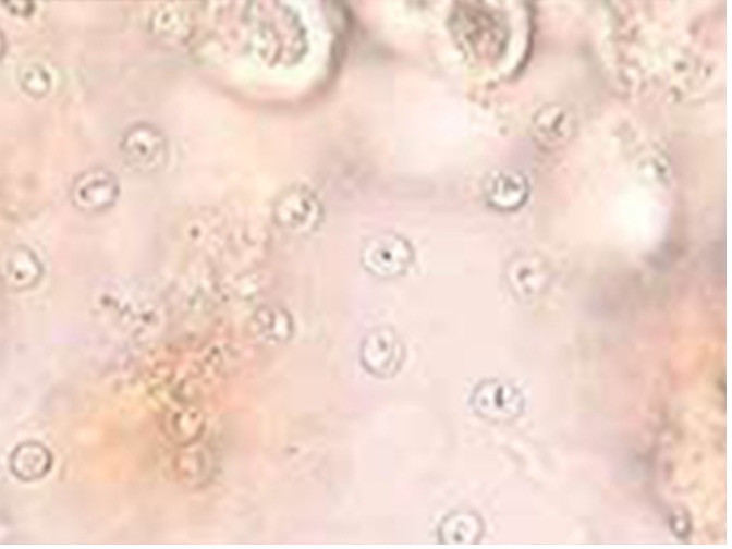
Oocysts of Cryptosporidium from fecal flotation
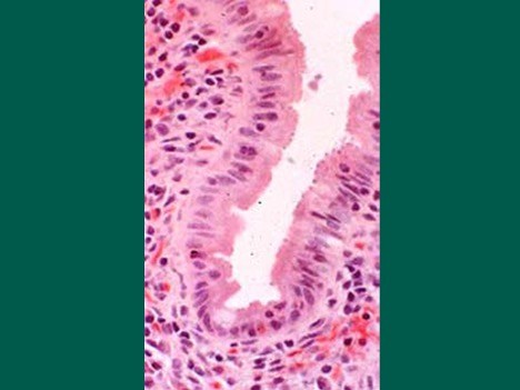
Photomicrograph of developmental stages of Cryptosporidium from the small intestine
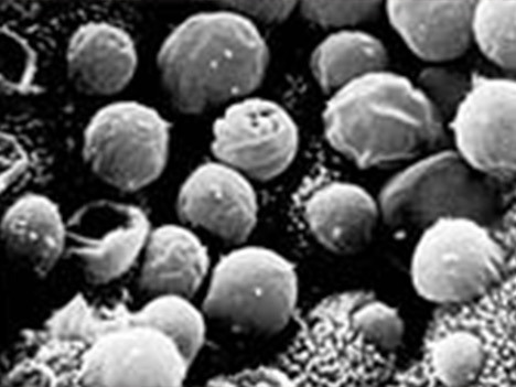
Scanning electron microscopy showing Cryptosporidium stages in the microvillus border of epithelial cells
Disease
- Cryptosporidiosis is a self-limited, small bowel infection characterized by secretory diarrhea. Fluid loss may be severe in clinical cases.
- The disease may be more severe, prolonged, and sometimes life-threatening in immunocompromised hosts.
- In many cases cats may be asymptomatic.
Prevalence
- Seroprevalence along with microscopic identification and PCR-based diagnostics studies indicate that Cryptosporidiuminfection is relatively common in cats.
- Confirmation of current infection by fecal examination is infrequent; commonly seen in only 1% to 5% of cases. Seroprevalence (antibody) studies, on the other hand, suggest higher rates of resolved infection; antibody titers are measurable in 20% to 50% of cases.
Transmission
- Infection occurs following ingestion of sporulated oocysts from fecal-contaminated environments, food, or water.
Host Associations and Transmission Between Hosts
- Cryptosporidium exists as many distinct species and genotypes with different degrees of infectivity for animals and humans:
- C. canis - dogs, rarely humans
- C. felis - cats, rarely humans
- C. hominis - only in humans
- C. parvum - a species in ruminants and other hosts that also will readily infect people.
- Although Cryptosporidium canis and Cryptosporidium felis primarily infect dogs and cats, respectively, zoonotic transmission to humans has been documented. See section on 'Zoonotic Potential of Valid Species and Genotypes of Cryptosporidium' for more information.
Prepatent Period and Environmental Factors
- Oocysts are generally shed in the feces 3 to 6 days after infection.
- Oocysts are immediately infectious when passed and are capable of surviving in the environment for extended periods.
- Oocysts are resistant to most disinfectants, including routine chlorine concentrations in drinking water and swimming pools.
- Oocysts are susceptible to commercial formulations of ammonia and heat over 70°C.
Site of Infection and Pathogenesis
- Cryptosporidium spp. infect the microvillar border of cells lining the small intestine and other organs. The developmental cycle is similar to Cystoisospora spp.
- In immunocompromised humans, infections may also occur in the large intestine, respiratory tract, biliary tract, pancreatic duct, and other sites.
- Most Cryptosporidium infections in cats are subclinical.
- Diarrhea and dehydration are the primary clinical signs
- In immunocompetent and healthy cats, the infections are usually self-limiting and some may be asymptomatic.
- Persistent infections usually denote an underlying cause.
Diagnosis
- Diagnosis can be difficult. Specimens should be sent to an academic or commercial testing laboratory familiar with the necessary diagnostic techniques.
- Specific species identification requires molecular methods (e.g., PCR) that are currently only performed by some reference laboratories as well as several academic laboratories.
- Oocysts are typically isolated using centrifugal flotation with sucrose.
- The oocysts are spheroid and small, 4-6 µm in diameter. In sucrose preparations, the oocysts float just under the coverslip on the slide
- Oocysts are clear or the walls may have a pinkish hue depending on the objective lens used.
- These are some of the smallest parasite diagnostic stages seen in fecal samples and require skill and practice for accurate diagnosis.
- Oocysts in acid fast stained fecal smears are red or pink.
- Fluorescent antibody tests (IFA) can be performed on fecal smears.
- Fecal antigen is detected with enzyme-linked immunosorbent assays (ELISAs).
- Fecal PCR tests for Cryptosporidium
- Commercial assays are available for detection of Cryptosporidium DNA.
- Nucleic acid is stable in the feces for 10 days at 4ºC.
- Both centrifugal fecal flotation and fecal PCR have their strengths and weaknesses, however, to ensure the widest breadth of detection of intestinal parasites in cats, fecal PCR tests should be combined with microscopic examination of feces.
- Commercial laboratories publish algorithms specific to their diagnostic tests and may be useful.
- Patient status and lifestyle are important considerations when interpreting Cryptosporidium positive diagnostic results.
Treatment
- Treatment is difficult and few drugs are consistently effective against Cryptosporidium.
- Symptomatic patients may require treatment, particularly if clinical signs are severe or persistent.
- Non-symptomatic patients may not require treatment.
- Patients living in a household with vulnerable or immunocompromised humans or pets should be treated.
- Ultimately, it is the practitioner's responsibility to use their best judgement in consultation with the owner in deciding treatment options.
- The following drugs have been used with some success in cases where animals have persistent diarrhea with oocyst shedding:
- Tylosin: 10 to 15 mg/kg TID for 14 to 21 days (cats)
- Azithromycin: 7 to 15 mg/kg for 5 to 7 days (cats)
- Until recently nitazoxanide was approved only for use in humans. While there are few studies in dogs, and none in cats, one study found that treatment with either 75 mg/kg or 150 mg/kg was effective in clearing Cryptosporidium spp. in dogs compared to controls.
- Diagnosing and treating any underlying immunosuppressive conditions is key to managing Cryptosporidium spp. in those patients that are ill.
- A key component to treatment may be normalizing the GI biome, which can be aided by probiotics and/or diets specifically formulated to support GI health.
Control and Prevention
- Due to the low infective dose and persistence of oocysts in the environment coupled with the lack of effective treatment, control and prevention should be focused on reducing environmental contamination with infectious oocyst and should consist of:
- Promptly picking up and removing feces to prevent fecal contamination of the environment
- Cleaning with heat or steam followed by disinfection and drying to promote desiccation, may be useful.
Public Health Considerations
- The zoonotic risk of all recognized Cryptosporidium species and genotypes is not known. However, 19 species and 4 genotypes have been identified in humans (See “Zoonotic Potential of Valid Species and Genotypes of Cryptosporidium” for more information).
- Human infections are most often due to C. hominis and C. parvum.
- Other species more commonly implicated in human infections are C. meleagridis, C. felis, C. canis, and C. ubiquitum. However, the risk for acquiring these Cryptosporidium species in comparison to C. hominis and C. parvum is much lower.
- In the United States, Cryptosporidium infections are the leading cause of waterborne illness in humans.
- To reduce the risk of acquiring Cryptosporidium infections, people should practice good hygiene, avoid swimming in potentially contaminated water, and avoid drinking untreated water or consuming potentially contaminated food.
Zoonotic Potential of Valid Species and Genotypes of Cryptosporidium
- There are 44 recognized species and numerous genotypes of Cryptosporidium.
- Of these, 19 species and 4 genotypes have been identified in human infections either from immunocompetent or immunocompromised individuals. Below is a summary of the Cryptosporidium species and genotypes found in human infections as well as their primary host species.
C. hominis
- Primary Host: Humans
C. parvum
- Primary Host: Many mammals
C. meleagridis
- Primary Host: Avians
C. canis
- Primary Host: Dogs
C. felis
- Primary Host: Cats
C. ubiquitum
- Primary Host: Many mammals
C. cuniculus
- Primary Host: Rabbits
C. viatorum
- Primary Host: Rodents
C. muris
- Primary Host: Rodents
C. andersoni
- Primary Host: Cattle
C. erinacei
- Primary Host: Hedgehogs
C. tyzzeri
- Primary Host: Rodents
C. bovis
- Primary Host: Cattle
C. suis
- Primary Host: Pigs
C. scrofarum
- Primary Host: Pigs
C. occultus
- Primary Host: Rodents
C. xiaoi
- Primary Host: Small ruminants
C. fayeri
- Primary Host: Marsupials
C. ditrichi
- Primary Host: Rodents
Chipmunk genotype I
- Primary Host: Rodents
Mink genotype
- Primary Host: Mink
Skunk genotype
- Primary Host: Skunk
Horse genotype
- Primary Host: Horse
Selected References
- Barbosa AD, Egan S, Feng Y, Xiao L, Ryan U. 2023. Cryptosporidium and Giardia in cats and dogs: What is the real zoonotic risk? Curr Res Parasitol Vector Borne Dis 4: Article 100158.
- Lucio-Forster A, et al, 2010. Minimal zoonotic risk of cryptosporidiosis from pet dogs and cats. Trends Parasitol., 6:4:174.
- Ryan U, Zahedi A, Feng Y, Xiao L. 2021. An update on zoonotic Cryptosporidium species and genotypes in humans. Animals (Basel) 11: Article 3307.
Synopsis
CAPC Recommends
- Continuing to monitor the health of the pet for those cats that test positive for Cryptosporidium spp. on annual wellness fecal testing.
- Diagnosing and treating any underlying conditions that may be weakening the animal’s immune system or causing stress, which can allow Cryptosporidium species to multiply.
- Using diet and supplements to support GI health and normalize the GI biome.
Introduction
- Cryptosporidium species are coccidian-like protozoan parasites that infect a wide variety of vertebrate hosts.
- Most species and strains are highly adapted to specific hosts.
- When other hosts are exposed to these strains, they are less likely to be infected unless they are immunocompromised.
- Humans are more likely to become infected by C. hominis and certain genotypes of C. parvum than by a feline species.
- However, some species are thought to be shared among dogs, cats, and humans.
Stages
- Sporulated oocysts—resistant stages for environmental transmission—are infective when shed by a host.
- Schizonts (asexual stages)
- Gametes (sexual stages)
Oocysts of Cryptosporidium from fecal flotation
Photomicrograph of developmental stages of Cryptosporidium from the small intestine
Scanning electron microscopy showing Cryptosporidium stages in the microvillus border of epithelial cells
Disease
- Cryptosporidiosis is a self-limited, small bowel infection characterized by secretory diarrhea. Fluid loss may be severe in clinical cases.
- The disease may be more severe, prolonged, and sometimes life-threatening in immunocompromised hosts.
- In many cases cats may be asymptomatic.
Prevalence
- Seroprevalence along with microscopic identification and PCR-based diagnostics studies indicate that Cryptosporidiuminfection is relatively common in cats.
- Confirmation of current infection by fecal examination is infrequent; commonly seen in only 1% to 5% of cases. Seroprevalence (antibody) studies, on the other hand, suggest higher rates of resolved infection; antibody titers are measurable in 20% to 50% of cases.
Transmission
- Infection occurs following ingestion of sporulated oocysts from fecal-contaminated environments, food, or water.
Host Associations and Transmission Between Hosts
- Cryptosporidium exists as many distinct species and genotypes with different degrees of infectivity for animals and humans:
- C. canis - dogs, rarely humans
- C. felis - cats, rarely humans
- C. hominis - only in humans
- C. parvum - a species in ruminants and other hosts that also will readily infect people.
- Although Cryptosporidium canis and Cryptosporidium felis primarily infect dogs and cats, respectively, zoonotic transmission to humans has been documented. See section on 'Zoonotic Potential of Valid Species and Genotypes of Cryptosporidium' for more information.
Prepatent Period and Environmental Factors
- Oocysts are generally shed in the feces 3 to 6 days after infection.
- Oocysts are immediately infectious when passed and are capable of surviving in the environment for extended periods.
- Oocysts are resistant to most disinfectants, including routine chlorine concentrations in drinking water and swimming pools.
- Oocysts are susceptible to commercial formulations of ammonia and heat over 70°C.
Site of Infection and Pathogenesis
- Cryptosporidium spp. infect the microvillar border of cells lining the small intestine and other organs. The developmental cycle is similar to Cystoisospora spp.
- In immunocompromised humans, infections may also occur in the large intestine, respiratory tract, biliary tract, pancreatic duct, and other sites.
- Most Cryptosporidium infections in cats are subclinical.
- Diarrhea and dehydration are the primary clinical signs
- In immunocompetent and healthy cats, the infections are usually self-limiting and some may be asymptomatic.
- Persistent infections usually denote an underlying cause.
Diagnosis
- Diagnosis can be difficult. Specimens should be sent to an academic or commercial testing laboratory familiar with the necessary diagnostic techniques.
- Specific species identification requires molecular methods (e.g., PCR) that are currently only performed by some reference laboratories as well as several academic laboratories.
- Oocysts are typically isolated using centrifugal flotation with sucrose.
- The oocysts are spheroid and small, 4-6 µm in diameter. In sucrose preparations, the oocysts float just under the coverslip on the slide
- Oocysts are clear or the walls may have a pinkish hue depending on the objective lens used.
- These are some of the smallest parasite diagnostic stages seen in fecal samples and require skill and practice for accurate diagnosis.
- Oocysts in acid fast stained fecal smears are red or pink.
- Fluorescent antibody tests (IFA) can be performed on fecal smears.
- Fecal antigen is detected with enzyme-linked immunosorbent assays (ELISAs).
- Fecal PCR tests for Cryptosporidium
- Commercial assays are available for detection of Cryptosporidium DNA.
- Nucleic acid is stable in the feces for 10 days at 4ºC.
- Both centrifugal fecal flotation and fecal PCR have their strengths and weaknesses, however, to ensure the widest breadth of detection of intestinal parasites in cats, fecal PCR tests should be combined with microscopic examination of feces.
- Commercial laboratories publish algorithms specific to their diagnostic tests and may be useful.
- Patient status and lifestyle are important considerations when interpreting Cryptosporidium positive diagnostic results.
Treatment
- Treatment is difficult and few drugs are consistently effective against Cryptosporidium.
- Symptomatic patients may require treatment, particularly if clinical signs are severe or persistent.
- Non-symptomatic patients may not require treatment.
- Patients living in a household with vulnerable or immunocompromised humans or pets should be treated.
- Ultimately, it is the practitioner's responsibility to use their best judgement in consultation with the owner in deciding treatment options.
- The following drugs have been used with some success in cases where animals have persistent diarrhea with oocyst shedding:
- Tylosin: 10 to 15 mg/kg TID for 14 to 21 days (cats)
- Azithromycin: 7 to 15 mg/kg for 5 to 7 days (cats)
- Until recently nitazoxanide was approved only for use in humans. While there are few studies in dogs, and none in cats, one study found that treatment with either 75 mg/kg or 150 mg/kg was effective in clearing Cryptosporidium spp. in dogs compared to controls.
- Diagnosing and treating any underlying immunosuppressive conditions is key to managing Cryptosporidium spp. in those patients that are ill.
- A key component to treatment may be normalizing the GI biome, which can be aided by probiotics and/or diets specifically formulated to support GI health.
Control and Prevention
- Due to the low infective dose and persistence of oocysts in the environment coupled with the lack of effective treatment, control and prevention should be focused on reducing environmental contamination with infectious oocyst and should consist of:
- Promptly picking up and removing feces to prevent fecal contamination of the environment
- Cleaning with heat or steam followed by disinfection and drying to promote desiccation, may be useful.
Public Health Considerations
- The zoonotic risk of all recognized Cryptosporidium species and genotypes is not known. However, 19 species and 4 genotypes have been identified in humans (See “Zoonotic Potential of Valid Species and Genotypes of Cryptosporidium” for more information).
- Human infections are most often due to C. hominis and C. parvum.
- Other species more commonly implicated in human infections are C. meleagridis, C. felis, C. canis, and C. ubiquitum. However, the risk for acquiring these Cryptosporidium species in comparison to C. hominis and C. parvum is much lower.
- In the United States, Cryptosporidium infections are the leading cause of waterborne illness in humans.
- To reduce the risk of acquiring Cryptosporidium infections, people should practice good hygiene, avoid swimming in potentially contaminated water, and avoid drinking untreated water or consuming potentially contaminated food.
Zoonotic Potential of Valid Species and Genotypes of Cryptosporidium
- There are 44 recognized species and numerous genotypes of Cryptosporidium.
- Of these, 19 species and 4 genotypes have been identified in human infections either from immunocompetent or immunocompromised individuals. Below is a summary of the Cryptosporidium species and genotypes found in human infections as well as their primary host species.
C. hominis
- Primary Host: Humans
C. parvum
- Primary Host: Many mammals
C. meleagridis
- Primary Host: Avians
C. canis
- Primary Host: Dogs
C. felis
- Primary Host: Cats
C. ubiquitum
- Primary Host: Many mammals
C. cuniculus
- Primary Host: Rabbits
C. viatorum
- Primary Host: Rodents
C. muris
- Primary Host: Rodents
C. andersoni
- Primary Host: Cattle
C. erinacei
- Primary Host: Hedgehogs
C. tyzzeri
- Primary Host: Rodents
C. bovis
- Primary Host: Cattle
C. suis
- Primary Host: Pigs
C. scrofarum
- Primary Host: Pigs
C. occultus
- Primary Host: Rodents
C. xiaoi
- Primary Host: Small ruminants
C. fayeri
- Primary Host: Marsupials
C. ditrichi
- Primary Host: Rodents
Chipmunk genotype I
- Primary Host: Rodents
Mink genotype
- Primary Host: Mink
Skunk genotype
- Primary Host: Skunk
Horse genotype
- Primary Host: Horse
Selected References
- Barbosa AD, Egan S, Feng Y, Xiao L, Ryan U. 2023. Cryptosporidium and Giardia in cats and dogs: What is the real zoonotic risk? Curr Res Parasitol Vector Borne Dis 4: Article 100158.
- Lucio-Forster A, et al, 2010. Minimal zoonotic risk of cryptosporidiosis from pet dogs and cats. Trends Parasitol., 6:4:174.
- Ryan U, Zahedi A, Feng Y, Xiao L. 2021. An update on zoonotic Cryptosporidium species and genotypes in humans. Animals (Basel) 11: Article 3307.