Mesocestoides
Mesocestoides for Dog Last updated: Mar 16, 2026
Synopsis
CAPC Recommends
- Treating intestinal infections with praziquantel to reduce environmental contamination
- Crushing the proglottid onto a microscope slide to visualize the clear, round eggs with the long distinct hooks
- Treatment of tetrathyridial stages is not always effective. Aggressive treatment (surgery/lavage) combined with long-term drug administration is recommended for the best prognosis. However, recurrences of infection is common.
- Peritoneal or pleural Mesocestoides spp. Infection should be considered as part of the differential diagnosis for any dog or cat presenting with nonspecific, progressive clinical signs combined with ascites or pleural effusion.
Species
Mesocestoides spp.
Overview of Life Cycle
- Mesocestoides spp. tapeworms have an indirect lifecycle that has not been fully elucidated. While the number of required intermediate hosts is debated, evidence suggests that they require at least two intermediate hosts. The first intermediate host is unknown, but many vertebrates can serve as a final intermediate host harboring larval tapeworms (tetrathyridium) within body cavities, including snakes, birds, lizards, rodents, cats and dogs. These tetrathyridia are able to replicate asexually within the intermediate host.
- Dogs and cats can be intermediate or definitive hosts for Mesocestoides spp. tapeworms, the latter of which shed egg-laden proglottids in their feces.
Stages
- The infectious egg contains a hexacanth embryo.
- Both cephalic and acephalic tetrathyridia (metacestode stage) are reported. The cephalic form has four suckers.
- Adult Mesocestoides spp. are found in the small intestine of an infected dog or cat, and proglottids are shed in the feces.

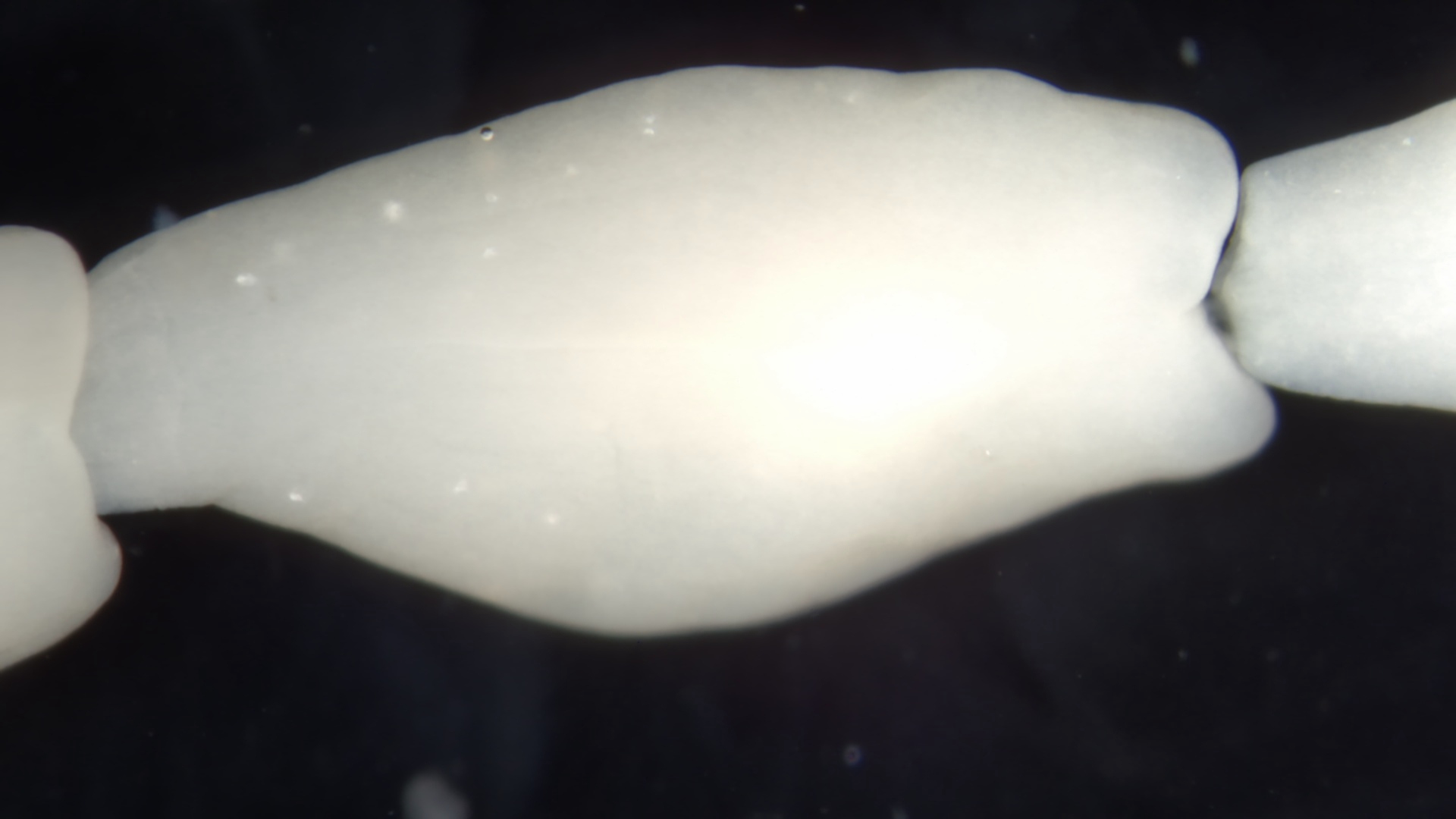
Mesocestoides spp. scolex

Mesocestoides spp. pore
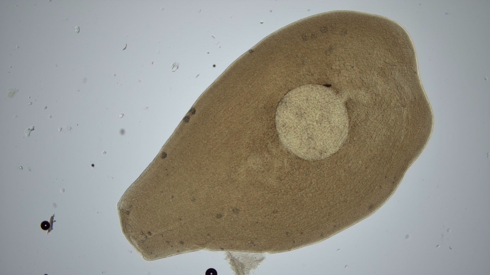
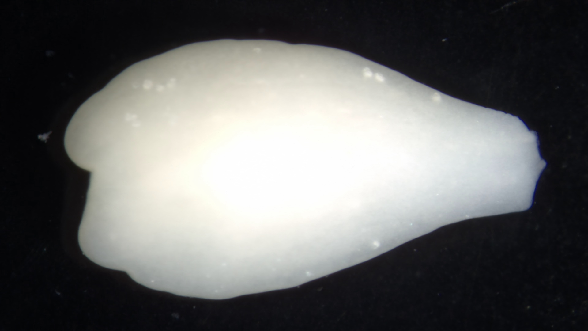
Mesocestoides proglottid
Mesocestoides chain
Mesocestoides single
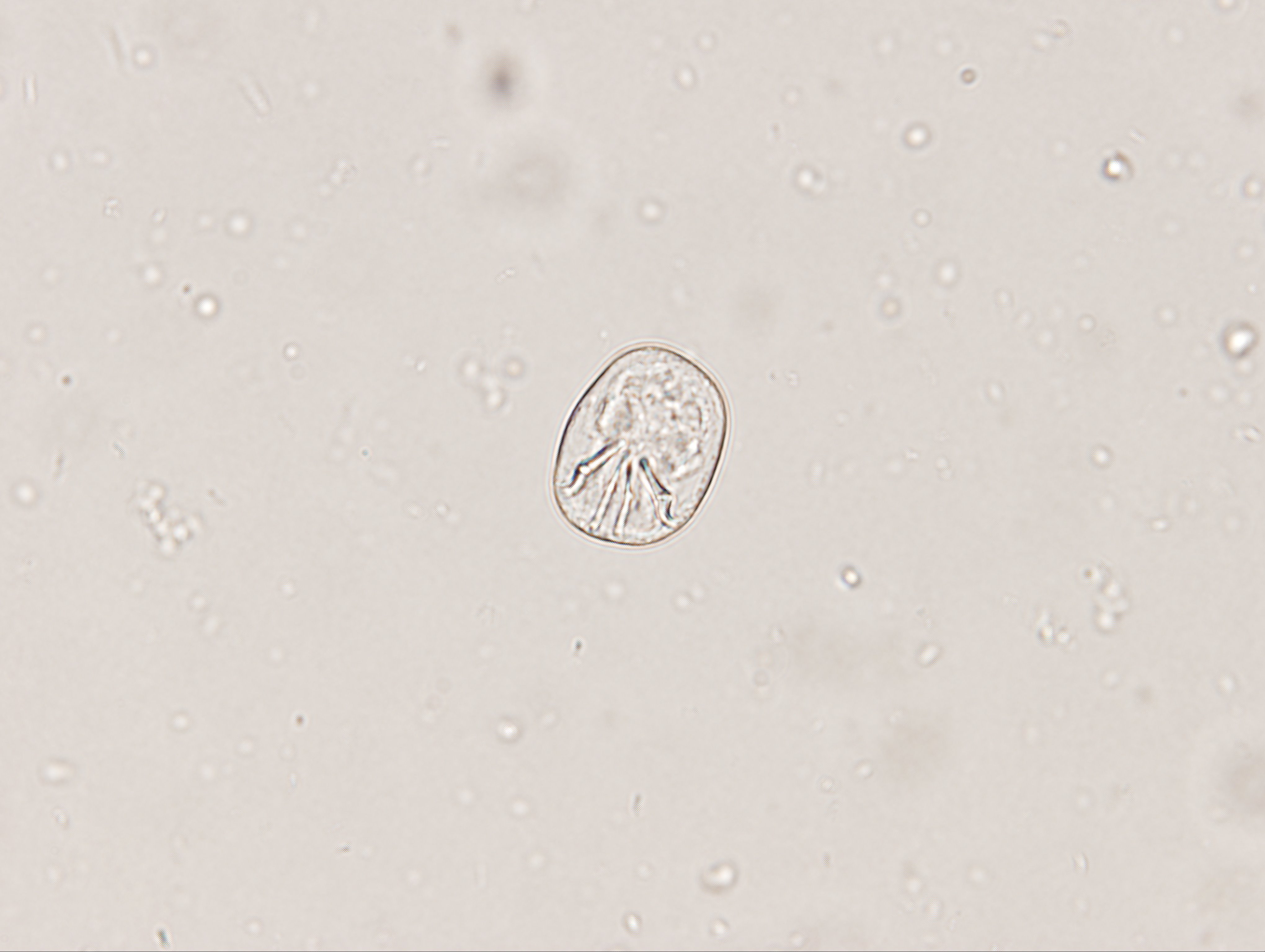
Mesocestoides egg
Disease
- Two main forms of disease are caused by infection of small animals with Mesocestoides spp.
- The intestinal form in which dogs and cats harbor adult Mesoscestoides spp. rarely causes clinical disease.
- Peritoneal mesocestoidiasis is a rare condition in which tetrathyridium asexually replicate within the peritoneal cavity, causing clinical signs such as ascites, anorexia/weight loss, vomiting, diarrhea, and tachypnea. Scrotal infections have also been documented, and scrotal swelling may be clinically visible.
- Peritoneal mesocestoidiasis may also be clinically silent and found incidentally upon opening the abdominal cavity for surgical procedures.
- Extremely rare, pleural mesocestoidiasis has also been described. Clinical signs may include pleural effusion, respiratory distress, and cyanosis.
Prevalence
- The prevalence of Mesocestoides spp. in dogs and cats in North America is reported to be as low as 0-1%. The global estimated prevalence based on pooled data in dogs and cats is approximately 8%. A number of factors influence the likelihood that a dog or cat will be infected with tapeworms, including the geographic region and the opportunity the animal may have to ingest an infected intermediate host.
- Prevalence data generated by fecal flotation alone underestimates the frequency of infection with Mesocestoides spp. because proglottids (and thus eggs) are focally distributed in fecal material and because eggs are heavy and thus do not readily float; a given fecal sample may be negative for tapeworm proglottids or eggs, even in the presence of an infection.
- Mesocestoides spp. are found throughout North America with higher incidence of reported infections in the western and southeastern United States.
- Peritoneal infections with Mesocestoides spp. have been most commonly reported in California and other western states in dogs.
Host Associations and Transmission Between Hosts
- Both dogs and cats are susceptible to infection with Mesocestoides spp. following ingestion of an infected intermediate host containing tetrathyridia.
- Both dogs and cats have been reported as second intermediate hosts in which tetrathyridia infect the peritoneal space. The route of exposure for development of this form of the disease is not known. While extremely rare, tetrathyridia have also been reported to infect the pleural cavity.
Prepatent Period and Environmental Factors
- Dogs and cats may begin shedding proglottids of Mesocestoides spp. as soon as 3 weeks following infection.
Site of Infection and Pathogenesis
Intestinal infection
- Adult tapeworms are found in the small intestine of dogs and cats. Motile proglottids may be seen in the perianal region as they exit the animal, in the pet’s environment (e.g., on bedding), or in the fecal material itself. Intestinal Mesocestoides spp. infections typically do not cause significant disease in dogs and cats, but because they are aesthetically unpleasant, treatment is warranted.
Peritoneal infection
- The metacestodes (tetrathyridia) are found within the peritoneal cavity of dogs and cats and have been reported in the scrotum of intact males. Tetrathyridia reproduce asexually, leading to dramatic expansion of the parasite. These infections can remain asymptomatic or can cause irritation within the abdominal cavity leading to inflammation, ascites, and abdominal distension. Other signs that may accompany the peritoneal form of Mesocestoides spp. infection include vomiting, anorexia, depression, diarrhea, tachypnea, and scrotal swelling.
Pleural infection
- The tetrathyridia are found within the pleural cavity of dogs and cats and reproduce asexually similarly to what is seen with peritoneal infections. These infections can cause pleuritis and pleural effusion. Accompanying signs may include those related to respiratory distress.
Diagnosis
Intestinal form
- Diagnosis of intestinal infection with tapeworms is reached by identifying proglottids in the fecal material or by recognizing eggs upon fecal flotation.
- However, because proglottids are not uniformly distributed in the fecal material and eggs do not consistently float, fecal flotation alone is not a reliable means of diagnosing tapeworm infection in dogs and cats.
- Proglottids can be put onto a microscope slide and crushed with a drop of water to release as visualize the eggs microscopically
- Eggs are thin-walled, measure approximately 30-40 microns, and contain a hexacanth embryo.
- Adult Mesocestoides spp. are 30-70cm long and have a scolex with 4 muscular suckers and no rostellum (hooks).
- Mature proglottids are 3-4mm in length and can be identified based on the presence of a parauterine organ, located on the midline of the proglottid, and a genital pore that opens ventrally.
Peritoneal form
- Diagnosis of peritoneal Mesocestoides spp. infection is based on clinical signs and diagnostic tools such as laparoscopic exam, cytology, radiographs, ultrasound, and PCR.
- Tetrathyridia are reported in two forms. The cephalic form can be identified by the presence of four suckers. The acephalic form can only be definitively identified by PCR. Both are small (100 microns to 3mm), motile, irregularly shaped organisms.
- Cytology of abdominal fluid may contain tetrathyridia and/or calcareous corpuscles, which are small refractile structures characterized by concentric rings.
- Radiography and ultrasonography may be used to support the diagnosis but do not reveal changes pathognomonic for peritoneal larval cestodiasis. Radiographic changes include increased radiopacity and ground-glass appearance, which are consistent with peritoneal effusion. Ultrasound studies have revealed anechoic cystic structures varying in complexity within the abdomen.
Pleural form
- Diagnosis of pleural Mesocestoides spp. infection is similar to those utilized for diagnosing peritoneal infections.
- Cytology of pleural effusion may contain tetrathyridia and/or calcareous corpuscles.
- Similar to the peritoneal form of the disease, radiography and ultrasonography may be used to support the diagnosis. Radiographic changes are often consistent with pleural effusion. Ultrasound findings may include pleural effusion characterized by corpusculate fluid and the presence of anechoic cystic structures.
Treatment
- Praziquantel at 5mg/kg can be given orally or subcutaneously for the treatment of Mesocestoides spp. adult tapeworm infections in the intestine of dogs and cats.
- Treatment of adult tapeworms in dogs and cats must be combined with appropriate management, such as prevention of ingestion of prey species; in the absence of these changes, re-infection may occur.
- Treatment of tetrathyridial stages of Mesocestoides spp. requires long-term drug administration and numerous follow up visits.
- Peritoneal lavage should be performed to remove as many tetrathyridia as possible.
- Fenbendazole, (100 mg/kg, twice daily for 28 days) while not labeled for the treatment of peritoneal Mesocestoides spp. infection, has been reported as curative in some dogs and is generally the recommended treatment protocol. This dose of fenbendazole has been associated with bone marrow suppression in dogs as well, so veterinarians should use caution and be prepared to provide supportive care if this protocol is initiated. However, combinations of fenbendazole and praziquantel or lower doses of fenbendazole have also been utilized with varying success.
- Long-term monitoring is needed after treatment because reoccurrence of infection is common, which is likely due to asexual reproduction of any remaining tetrathyridia.
- Dogs and cats that become persistently infected with Mesocestoides spp. may be given fenbendazole (50-100mg/kg) daily for the life of the animal. Caution should be used if giving high doses of fenbendazole, see above.
- The prognosis for the peritoneal form of Mesocestoides spp. infection is guarded.
- Treatment for pleural infections has been successful using high doses of fenbendazole. For severe disease, this may need to be combined with surgery and/or lavage.
Control and Prevention
- In dogs or cats that are allowed outside or that are known to have predatory behavior, a heartworm preventive containing praziquantel would be expected to help control intestinal Mesocestoides spp. infections.
- Prevention of predation and scavenging activity by keeping cats indoors and dogs confined to a leash or in a fenced yard will limit the opportunity for dogs and cats to acquire infection with Mesocestoides spp. via ingestion of tetrathyridia in intermediate hosts.
- The life cycle of this parasite is not fully understood, which precludes recommendations regarding specific preventive measures to avoid peritoneal or pleural infection with Mesocestoides spp. tapeworms.
Public Health Considerations
- Dogs and cats that shed gravid proglottids do not pose a direct zoonotic risk as Mesocestoides spp. eggs are not infective to humans.
- Human infection with Mesocestoides spp. is uncommon, and intestinal infections are usually well tolerated and easily treated. These infections are usually linked to ingestion of improperly prepared food, such as raw or undercooked viscera or blood that contains tetrathyridia.
Selected References
- Bonfanti U, et al. 2004. Clinical, cytological and molecular evidence of Mesocestoides spp. infection in a dog from Italy. J Vet Med A 51:435-438.
- Boyce W, et al. 2011. Survival analysis of dogs diagnosed with canine peritoneal larval cestodiasis (Mesocestoides spp.). Vet Parasit 180: 256-261.
- Carta S, et al. 2021. Clinical forms of peritoneal larval cestodiasis by Mesocestoides spp. in dogs: diagnosis, treatment and long term follow-up. Parasitol Res 120:1727-1735.
- Conboy G. 2012. Cestodes of dogs and cats in North America. Vet Clin North Am Small Anim Pract 39:1075-1090.
- Jesudoss Chelladurai JRJ, Brewer MT. 2021. Global prevalence of Mesocestoides infections in animals – A systematic review and meta-analysis. Vet Parasit 298:109537.
- McAllister CT, et al. 2018. Morphological and molecular characterization of post-larval pre-tetrathyridia of Mesocestoides sp. (Cestoda: Cyclophyllidea) from Gound Skin, Scincella lateralis (Sauria: Scincidae), from Southeastern Oklahoma. J Parasitol 104:246-253.
- Petrescu V, et al. 2020. Severe pleural effusion in a dog affected by larval mesocestodiasis. Top Companion Anim Med 40:100450.
- Sindičić M, et al. 2021. First description of peritoneal and pleural metacestodosis caused by Mesocestoides vogae in a European wild cat (Felis silvestris silvestris). Parasitol Res 120:2275-2279.
Synopsis
CAPC Recommends
- Treating intestinal infections with praziquantel to reduce environmental contamination
- Crushing the proglottid onto a microscope slide to visualize the clear, round eggs with the long distinct hooks
- Treatment of tetrathyridial stages is not always effective. Aggressive treatment (surgery/lavage) combined with long-term drug administration is recommended for the best prognosis. However, recurrences of infection is common.
- Peritoneal or pleural Mesocestoides spp. Infection should be considered as part of the differential diagnosis for any dog or cat presenting with nonspecific, progressive clinical signs combined with ascites or pleural effusion.
Species
Mesocestoides spp.
Overview of Life Cycle
- Mesocestoides spp. tapeworms have an indirect lifecycle that has not been fully elucidated. While the number of required intermediate hosts is debated, evidence suggests that they require at least two intermediate hosts. The first intermediate host is unknown, but many vertebrates can serve as a final intermediate host harboring larval tapeworms (tetrathyridium) within body cavities, including snakes, birds, lizards, rodents, cats and dogs. These tetrathyridia are able to replicate asexually within the intermediate host.
- Dogs and cats can be intermediate or definitive hosts for Mesocestoides spp. tapeworms, the latter of which shed egg-laden proglottids in their feces.
Stages
- The infectious egg contains a hexacanth embryo.
- Both cephalic and acephalic tetrathyridia (metacestode stage) are reported. The cephalic form has four suckers.
- Adult Mesocestoides spp. are found in the small intestine of an infected dog or cat, and proglottids are shed in the feces.
Mesocestoides spp. scolex
Mesocestoides spp. pore
Mesocestoides proglottid
Mesocestoides chain
Mesocestoides single
Mesocestoides egg
Disease
- Two main forms of disease are caused by infection of small animals with Mesocestoides spp.
- The intestinal form in which dogs and cats harbor adult Mesoscestoides spp. rarely causes clinical disease.
- Peritoneal mesocestoidiasis is a rare condition in which tetrathyridium asexually replicate within the peritoneal cavity, causing clinical signs such as ascites, anorexia/weight loss, vomiting, diarrhea, and tachypnea. Scrotal infections have also been documented, and scrotal swelling may be clinically visible.
- Peritoneal mesocestoidiasis may also be clinically silent and found incidentally upon opening the abdominal cavity for surgical procedures.
- Extremely rare, pleural mesocestoidiasis has also been described. Clinical signs may include pleural effusion, respiratory distress, and cyanosis.
Prevalence
- The prevalence of Mesocestoides spp. in dogs and cats in North America is reported to be as low as 0-1%. The global estimated prevalence based on pooled data in dogs and cats is approximately 8%. A number of factors influence the likelihood that a dog or cat will be infected with tapeworms, including the geographic region and the opportunity the animal may have to ingest an infected intermediate host.
- Prevalence data generated by fecal flotation alone underestimates the frequency of infection with Mesocestoides spp. because proglottids (and thus eggs) are focally distributed in fecal material and because eggs are heavy and thus do not readily float; a given fecal sample may be negative for tapeworm proglottids or eggs, even in the presence of an infection.
- Mesocestoides spp. are found throughout North America with higher incidence of reported infections in the western and southeastern United States.
- Peritoneal infections with Mesocestoides spp. have been most commonly reported in California and other western states in dogs.
Host Associations and Transmission Between Hosts
- Both dogs and cats are susceptible to infection with Mesocestoides spp. following ingestion of an infected intermediate host containing tetrathyridia.
- Both dogs and cats have been reported as second intermediate hosts in which tetrathyridia infect the peritoneal space. The route of exposure for development of this form of the disease is not known. While extremely rare, tetrathyridia have also been reported to infect the pleural cavity.
Prepatent Period and Environmental Factors
- Dogs and cats may begin shedding proglottids of Mesocestoides spp. as soon as 3 weeks following infection.
Site of Infection and Pathogenesis
Intestinal infection
- Adult tapeworms are found in the small intestine of dogs and cats. Motile proglottids may be seen in the perianal region as they exit the animal, in the pet’s environment (e.g., on bedding), or in the fecal material itself. Intestinal Mesocestoides spp. infections typically do not cause significant disease in dogs and cats, but because they are aesthetically unpleasant, treatment is warranted.
Peritoneal infection
- The metacestodes (tetrathyridia) are found within the peritoneal cavity of dogs and cats and have been reported in the scrotum of intact males. Tetrathyridia reproduce asexually, leading to dramatic expansion of the parasite. These infections can remain asymptomatic or can cause irritation within the abdominal cavity leading to inflammation, ascites, and abdominal distension. Other signs that may accompany the peritoneal form of Mesocestoides spp. infection include vomiting, anorexia, depression, diarrhea, tachypnea, and scrotal swelling.
Pleural infection
- The tetrathyridia are found within the pleural cavity of dogs and cats and reproduce asexually similarly to what is seen with peritoneal infections. These infections can cause pleuritis and pleural effusion. Accompanying signs may include those related to respiratory distress.
Diagnosis
Intestinal form
- Diagnosis of intestinal infection with tapeworms is reached by identifying proglottids in the fecal material or by recognizing eggs upon fecal flotation.
- However, because proglottids are not uniformly distributed in the fecal material and eggs do not consistently float, fecal flotation alone is not a reliable means of diagnosing tapeworm infection in dogs and cats.
- Proglottids can be put onto a microscope slide and crushed with a drop of water to release as visualize the eggs microscopically
- Eggs are thin-walled, measure approximately 30-40 microns, and contain a hexacanth embryo.
- Adult Mesocestoides spp. are 30-70cm long and have a scolex with 4 muscular suckers and no rostellum (hooks).
- Mature proglottids are 3-4mm in length and can be identified based on the presence of a parauterine organ, located on the midline of the proglottid, and a genital pore that opens ventrally.
Peritoneal form
- Diagnosis of peritoneal Mesocestoides spp. infection is based on clinical signs and diagnostic tools such as laparoscopic exam, cytology, radiographs, ultrasound, and PCR.
- Tetrathyridia are reported in two forms. The cephalic form can be identified by the presence of four suckers. The acephalic form can only be definitively identified by PCR. Both are small (100 microns to 3mm), motile, irregularly shaped organisms.
- Cytology of abdominal fluid may contain tetrathyridia and/or calcareous corpuscles, which are small refractile structures characterized by concentric rings.
- Radiography and ultrasonography may be used to support the diagnosis but do not reveal changes pathognomonic for peritoneal larval cestodiasis. Radiographic changes include increased radiopacity and ground-glass appearance, which are consistent with peritoneal effusion. Ultrasound studies have revealed anechoic cystic structures varying in complexity within the abdomen.
Pleural form
- Diagnosis of pleural Mesocestoides spp. infection is similar to those utilized for diagnosing peritoneal infections.
- Cytology of pleural effusion may contain tetrathyridia and/or calcareous corpuscles.
- Similar to the peritoneal form of the disease, radiography and ultrasonography may be used to support the diagnosis. Radiographic changes are often consistent with pleural effusion. Ultrasound findings may include pleural effusion characterized by corpusculate fluid and the presence of anechoic cystic structures.
Treatment
- Praziquantel at 5mg/kg can be given orally or subcutaneously for the treatment of Mesocestoides spp. adult tapeworm infections in the intestine of dogs and cats.
- Treatment of adult tapeworms in dogs and cats must be combined with appropriate management, such as prevention of ingestion of prey species; in the absence of these changes, re-infection may occur.
- Treatment of tetrathyridial stages of Mesocestoides spp. requires long-term drug administration and numerous follow up visits.
- Peritoneal lavage should be performed to remove as many tetrathyridia as possible.
- Fenbendazole, (100 mg/kg, twice daily for 28 days) while not labeled for the treatment of peritoneal Mesocestoides spp. infection, has been reported as curative in some dogs and is generally the recommended treatment protocol. This dose of fenbendazole has been associated with bone marrow suppression in dogs as well, so veterinarians should use caution and be prepared to provide supportive care if this protocol is initiated. However, combinations of fenbendazole and praziquantel or lower doses of fenbendazole have also been utilized with varying success.
- Long-term monitoring is needed after treatment because reoccurrence of infection is common, which is likely due to asexual reproduction of any remaining tetrathyridia.
- Dogs and cats that become persistently infected with Mesocestoides spp. may be given fenbendazole (50-100mg/kg) daily for the life of the animal. Caution should be used if giving high doses of fenbendazole, see above.
- The prognosis for the peritoneal form of Mesocestoides spp. infection is guarded.
- Treatment for pleural infections has been successful using high doses of fenbendazole. For severe disease, this may need to be combined with surgery and/or lavage.
Control and Prevention
- In dogs or cats that are allowed outside or that are known to have predatory behavior, a heartworm preventive containing praziquantel would be expected to help control intestinal Mesocestoides spp. infections.
- Prevention of predation and scavenging activity by keeping cats indoors and dogs confined to a leash or in a fenced yard will limit the opportunity for dogs and cats to acquire infection with Mesocestoides spp. via ingestion of tetrathyridia in intermediate hosts.
- The life cycle of this parasite is not fully understood, which precludes recommendations regarding specific preventive measures to avoid peritoneal or pleural infection with Mesocestoides spp. tapeworms.
Public Health Considerations
- Dogs and cats that shed gravid proglottids do not pose a direct zoonotic risk as Mesocestoides spp. eggs are not infective to humans.
- Human infection with Mesocestoides spp. is uncommon, and intestinal infections are usually well tolerated and easily treated. These infections are usually linked to ingestion of improperly prepared food, such as raw or undercooked viscera or blood that contains tetrathyridia.
Selected References
- Bonfanti U, et al. 2004. Clinical, cytological and molecular evidence of Mesocestoides spp. infection in a dog from Italy. J Vet Med A 51:435-438.
- Boyce W, et al. 2011. Survival analysis of dogs diagnosed with canine peritoneal larval cestodiasis (Mesocestoides spp.). Vet Parasit 180: 256-261.
- Carta S, et al. 2021. Clinical forms of peritoneal larval cestodiasis by Mesocestoides spp. in dogs: diagnosis, treatment and long term follow-up. Parasitol Res 120:1727-1735.
- Conboy G. 2012. Cestodes of dogs and cats in North America. Vet Clin North Am Small Anim Pract 39:1075-1090.
- Jesudoss Chelladurai JRJ, Brewer MT. 2021. Global prevalence of Mesocestoides infections in animals – A systematic review and meta-analysis. Vet Parasit 298:109537.
- McAllister CT, et al. 2018. Morphological and molecular characterization of post-larval pre-tetrathyridia of Mesocestoides sp. (Cestoda: Cyclophyllidea) from Gound Skin, Scincella lateralis (Sauria: Scincidae), from Southeastern Oklahoma. J Parasitol 104:246-253.
- Petrescu V, et al. 2020. Severe pleural effusion in a dog affected by larval mesocestodiasis. Top Companion Anim Med 40:100450.
- Sindičić M, et al. 2021. First description of peritoneal and pleural metacestodosis caused by Mesocestoides vogae in a European wild cat (Felis silvestris silvestris). Parasitol Res 120:2275-2279.