Dracunculus insignis
Synopsis
CAPC Recommends
- Dogs or cats with subcutaneous masses or emerging worms should be evaluated to determine if they are infected with Dracunculus insignis.
- Dogs and cats should not be allowed to ingest amphibian paratenic hosts or other possible paratenic hosts such as fish.
- Suspected worms should be preserved in 70-95% ethanol for identification because other worm species can be found in subcutaneous tissues.
Species
Canine and Feline
Dracunculus insignis is a parasite of numerous wild carnivore species (e.g., raccoons, fisher, mink, muskrat, opossums, fox, coyote, otter) but is occasionally found in dogs and cats. A related species, Dracunculus lutrae, has only been reported in otters.
Overview of Life Cycle
- Dogs, cats and wildlife hosts become infected with D. insignis via ingestion of aquatic copepods infected with L3 larvae. When ingested these larvae migrate to subcutaneous tissues where they mature and mate. After mating, females typically migrate to the legs where they become gravid. Females will induce the formation of a focal swelling that may ulcerate. When this lesion comes into contact with water, the female will release large numbers of L1 larvae. These larvae are consumed by copepods and develop to the L3 stage in 2-3 weeks.
- The complete life cycle can take 10-14 months.
- An additional possible transmission route is via ingestion of infected paratenic hosts (tadpoles, frogs, and possibly fish).
- Movie below shows copepod infected with several Dracunculus L3 larvae.
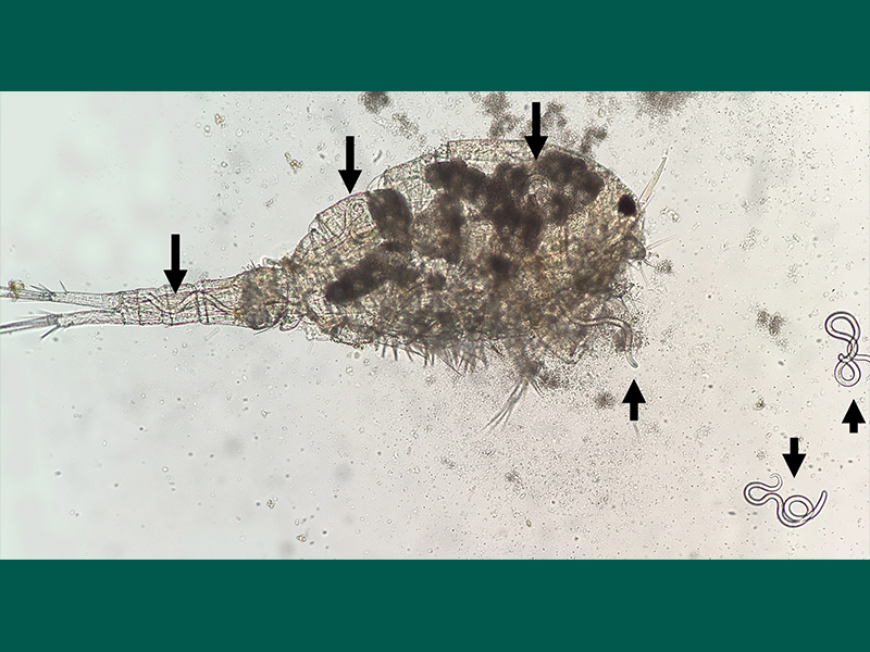
A copepod infected with Dracunculus (L3 larvae marked with arrows)
Stages
- Adult female worms are detected in the subcutaneous tissues or emerging from skin of dogs and cats. Male worms typically do not migrate to the extremities so are rarely observed. Female worms are large (17-31cm) while males, which are rarely found, are only 3-4cm long. Figure 2 shows an adult female extracted from naturally infected raccoon. Figure 3 shows an adult female Dracunculus emerging from the leg of a domestic dog.
- Larvae measure ~500µm x 50µm and have long pointed tails. They may be detected if a female worm is removed and placed in water (Figure 4) or in aspirates of masses. Absence of larvae does not rule out Dracunculus because females may not be fertile or gravid.

Adult female Dracunculus extracted from naturally infected raccoon

Adult female Dracunculus emerging from the leg of a domestic dog

L1 larvae removed from the uterus of an adult female D. insignis
Disease
- Cases in dogs and cats are rarely reported and are generally only detected right before or while the gravid female is emerging or has produced a grossly visible swelling.
- Adult female D. insignis may produce swelling and inflammation in the subcutaneous tissues and muscle fascia where present. These swellings may or may not be painful and may also be pruritic leading to localized alopecia. These generally are on the lower portions of the limbs but may occur anywhere on the body (e.g., thoracic wall, abdominal wall, and rarely females may emerge from the face or may be found in the eye).
- Following protrusion of gravid female worms through the skin to expel the larvae, the worm may be observed and there may be focal ulcerations of the skin followed by scarring. Figure 5 shows a post-emergent scar on a raccoon.

Post-emergent scar on a raccoon
Prevalence
- Diagnosed canine cases of D. insignis are uncommon and only a few feline cases have been reported. Canine cases have been reported throughout the eastern half of the United States and the cat cases were from New York, Alabama, Texas, and North Carolina. However, not all females emerge or cause swellings and males do not cause gross lesions so prevalence of infection in dogs and cats is likely higher. One study on ferrets infected with D. insignis found that only about 13% of 153 mature females emerged.
- Infections in dogs have been reported throughout the year; however, most reported cases in a retrospective study were diagnosed during the spring and winter months. The few documented cases in cats were reported during the same time frame.
- Infections in various wildlife species have been reported from numerous states in the eastern United States and Ontario, Canada. In some areas, infection prevalence in raccoons may exceed 50% when examined during the time of year (i.e., early-late spring) that females are becoming gravid and/or are emerging.
Host Associations and Transmission Between Hosts
- Dogs and cats become infected with Dracunculus upon ingestion of infected copepods so transmission is tied to aquatic environments.
- Amphibians and possibly other paratenic hosts (e.g. fish) can harbor infective L3 larvae.
- The parasite is not directly transmitted from an infected wildlife host to a dog or cat.
- A related parasite, the human guinea worm (D. medinensis) infects both humans and dogs in Africa, but there are no known human cases in North America caused by D. insignis.
Prepatent Period and Environmental Factors
- The prepatent period of D. insignis in dogs or cats is not known. However, data from raccoons and ferrets (experimental host) indicate that it takes 10-14 months for female worms to emerge and release L1 larvae.
- Infections in dogs less than 1 year of age should not be ruled out as there have been reported cases in dogs as young as 7 months.
Site of Infection and Pathogenesis
- Upon infection, larvae of D. insignis migrate out of the gastrointestinal tract and develop in subcutaneous tissues. No clinical signs or lesions are generally observed during this time.
- As female worms develop, they migrate subcutaneously to the site where they will emerge (most often a limb). During this migration, some worms may be deep within the muscle fascia which can cause pain and tenderness.
- Once fully gravid, female worms will produce a subcutaneous swelling that may ulcerate due to the release of uterine fluid that elicits an immune response.
Diagnosis
- Surgically extracted female worms from masses can only be identified to the genus level using morphologic criteria. For a definitive diagnosis, worms should be preserved in 70-95% ethanol for molecular identification.
- Male worms may be found in subcutaneous tissues at necropsy and can be identified using morphologic criteria. However, male worms are rarely identified due to their small size and lack of emergence from subcutaneous tissues.
- L1 larvae may be detected in aspirates of the swelling if females are gravid and if the needle punctures the worm.
- Several other parasites may be found in subcutaneous tissues of dogs or cats and must be differentiated from Dracunculus.
- Dirofilaria immitis, heartworm. On rare occasions D. immitis may undergo aberrant migration and be found in subcutaneous tissues. These worms can be distinguished from Dracunculus spp. morphologically or histologically (D. immitis have microfilariae whereas Dracunculus have larvae with long tails).
- Spirometra spp., plerocercoids. These tapeworms are easily distinguished morphologically and histologically from Dracunculus. These plerocercoids are much smaller than Dracunculus females and lack the thick cuticle of Dracunculus.
- Acanthocheilonema (Dipetalonema) reconditum, a filarial worm, resides in the subcutaneous tissues of dogs; however, adults of this species are very small (1.5-2.5 cm) and do not cause masses.

Coiled Spirometra in the subcutaneous tissues of a North American otter

Coiled Spirometra removed from the 'cyst' in the subcutaneous tissues of a North American otter
Treatment
- Surgical excision of female worms is the recommended treatment. Care should be taken to remove the worms intact. If a worm breaks it may elicit a severe inflammatory reaction or other complications if not removed promptly.
- Dogs and cats may benefit from antimicrobials to prevent secondary bacterial infections post-removal. Anti-inflammatories and antihistamines may also be of benefit to lessen the immune response. These ancillary therapies are more crucial in cases of incomplete removal of D. insignis from a subcutaneous mass or swelling.
- Various anthelmintic compounds (i.e., diethylcarbamazine, albendazole, ivermectin and, fenbendazole) have been evaluated and none were effective in decreasing worm burdens or killing adult female worms.
Control and Prevention
- Copepods are found in most natural bodies of water. It is currently unknown how commonly dogs and cats acquire infection from ingesting copepods during the act of drinking or if they acquire infections from ingesting amphibian or other possible paratenic hosts.
- Dogs and cats should not be allowed to ingest amphibian paratenic hosts or other possible paratenic hosts (e.g. fish).
Public Health Considerations
- D. insignis is not known to infect people; however, experimental exposure of a rhesus macaque (Macaca mulatta) resulted in infection with adult gravid female worms.
- Although infected dogs or cats indicate that the parasite is circulating in an area, these infected animals pose no risk to people or other pets in the household
- A related parasite, Dracunculus medinensis, is the human guinea worm and was once responsible for over 3 million cases annually in numerous countries in Africa and Asia. Beginning in the 1980s, the CDC and The Carter Center have led the efforts to eradicate this parasite using filters to remove copepods from drinking water (https://www.cartercenter.org/health/guinea_worm/index.html). These efforts have decreased the number to only 15 new human cases in two countries (Chad and South Sudan) during 2024. Infections in dogs have decreased in recent years, however, they present an ongoing challenge for eradication efforts. The highest reported number of D. medinensis infections in animals in 2024 occurred in Cameroon and Chad.
Selected References
- Beyer TA, Pinckney RD, Cooley AJ. Massive Dracunculus insignis infection in a dog. J Am Vet Med Assoc. 1999 Feb 1;214(3):366-8, 351.
- Cleveland CA, Eberhard ML, Thompson AT, Smith SJ, Zirimwabagabo H, Bringolf R, Yabsley MJ. Possible role of fish as transport hosts for Dracunculus spp. larvae. Emerg Infect Dis. 2017;23:1590-1592.
- Lucio-Forster A, Eberhard ML, Cama VA, Jenks MH, Jones C, Sanders SY, Pongratz JP, Bowman DD. First report of Dracunculus insignis in two naturally infected cats from the northeastern USA. J Feline Med Surg. 2014 Feb;16(2):194-7.
- Williams BM, Cleveland CA, Verocai GG, Swanepoel L, Niedringhaus KD, Paras KL, Nagamori Y, Little SE, Varela-Stokes A, Nemeth N, Wyrosdick H, Tucker A, Deal L, Gauthier D, Prouty S, DeAngelo C, Marsh A, Piepgras D, Cook LH, Milliren KB, Becker JS, Lyons C, Clark J, Stumph J, Borst MM, Craig T, Tucker KL, Ward A, Baird EM, Burke KA, Camp JW, Davis CA, Pulaski CN, Yabsley MJ. Dracunculus infections in domestic dogs and cats in North America; an under-recognized parasite? Vet Parasitol Reg Stud Reports. 2018;13:148-155.
Synopsis
CAPC Recommends
- Dogs or cats with subcutaneous masses or emerging worms should be evaluated to determine if they are infected with Dracunculus insignis.
- Dogs and cats should not be allowed to ingest amphibian paratenic hosts or other possible paratenic hosts such as fish.
- Suspected worms should be preserved in 70-95% ethanol for identification because other worm species can be found in subcutaneous tissues.
Species
Canine and Feline
Dracunculus insignis is a parasite of numerous wild carnivore species (e.g., raccoons, fisher, mink, muskrat, opossums, fox, coyote, otter) but is occasionally found in dogs and cats. A related species, Dracunculus lutrae, has only been reported in otters.
Overview of Life Cycle
- Dogs, cats and wildlife hosts become infected with D. insignis via ingestion of aquatic copepods infected with L3 larvae. When ingested these larvae migrate to subcutaneous tissues where they mature and mate. After mating, females typically migrate to the legs where they become gravid. Females will induce the formation of a focal swelling that may ulcerate. When this lesion comes into contact with water, the female will release large numbers of L1 larvae. These larvae are consumed by copepods and develop to the L3 stage in 2-3 weeks.
- The complete life cycle can take 10-14 months.
- An additional possible transmission route is via ingestion of infected paratenic hosts (tadpoles, frogs, and possibly fish).
- Movie below shows copepod infected with several Dracunculus L3 larvae.
A copepod infected with Dracunculus (L3 larvae marked with arrows)
Stages
- Adult female worms are detected in the subcutaneous tissues or emerging from skin of dogs and cats. Male worms typically do not migrate to the extremities so are rarely observed. Female worms are large (17-31cm) while males, which are rarely found, are only 3-4cm long. Figure 2 shows an adult female extracted from naturally infected raccoon. Figure 3 shows an adult female Dracunculus emerging from the leg of a domestic dog.
- Larvae measure ~500µm x 50µm and have long pointed tails. They may be detected if a female worm is removed and placed in water (Figure 4) or in aspirates of masses. Absence of larvae does not rule out Dracunculus because females may not be fertile or gravid.
Adult female Dracunculus extracted from naturally infected raccoon
Adult female Dracunculus emerging from the leg of a domestic dog
L1 larvae removed from the uterus of an adult female D. insignis
Disease
- Cases in dogs and cats are rarely reported and are generally only detected right before or while the gravid female is emerging or has produced a grossly visible swelling.
- Adult female D. insignis may produce swelling and inflammation in the subcutaneous tissues and muscle fascia where present. These swellings may or may not be painful and may also be pruritic leading to localized alopecia. These generally are on the lower portions of the limbs but may occur anywhere on the body (e.g., thoracic wall, abdominal wall, and rarely females may emerge from the face or may be found in the eye).
- Following protrusion of gravid female worms through the skin to expel the larvae, the worm may be observed and there may be focal ulcerations of the skin followed by scarring. Figure 5 shows a post-emergent scar on a raccoon.
Post-emergent scar on a raccoon
Prevalence
- Diagnosed canine cases of D. insignis are uncommon and only a few feline cases have been reported. Canine cases have been reported throughout the eastern half of the United States and the cat cases were from New York, Alabama, Texas, and North Carolina. However, not all females emerge or cause swellings and males do not cause gross lesions so prevalence of infection in dogs and cats is likely higher. One study on ferrets infected with D. insignis found that only about 13% of 153 mature females emerged.
- Infections in dogs have been reported throughout the year; however, most reported cases in a retrospective study were diagnosed during the spring and winter months. The few documented cases in cats were reported during the same time frame.
- Infections in various wildlife species have been reported from numerous states in the eastern United States and Ontario, Canada. In some areas, infection prevalence in raccoons may exceed 50% when examined during the time of year (i.e., early-late spring) that females are becoming gravid and/or are emerging.
Host Associations and Transmission Between Hosts
- Dogs and cats become infected with Dracunculus upon ingestion of infected copepods so transmission is tied to aquatic environments.
- Amphibians and possibly other paratenic hosts (e.g. fish) can harbor infective L3 larvae.
- The parasite is not directly transmitted from an infected wildlife host to a dog or cat.
- A related parasite, the human guinea worm (D. medinensis) infects both humans and dogs in Africa, but there are no known human cases in North America caused by D. insignis.
Prepatent Period and Environmental Factors
- The prepatent period of D. insignis in dogs or cats is not known. However, data from raccoons and ferrets (experimental host) indicate that it takes 10-14 months for female worms to emerge and release L1 larvae.
- Infections in dogs less than 1 year of age should not be ruled out as there have been reported cases in dogs as young as 7 months.
Site of Infection and Pathogenesis
- Upon infection, larvae of D. insignis migrate out of the gastrointestinal tract and develop in subcutaneous tissues. No clinical signs or lesions are generally observed during this time.
- As female worms develop, they migrate subcutaneously to the site where they will emerge (most often a limb). During this migration, some worms may be deep within the muscle fascia which can cause pain and tenderness.
- Once fully gravid, female worms will produce a subcutaneous swelling that may ulcerate due to the release of uterine fluid that elicits an immune response.
Diagnosis
- Surgically extracted female worms from masses can only be identified to the genus level using morphologic criteria. For a definitive diagnosis, worms should be preserved in 70-95% ethanol for molecular identification.
- Male worms may be found in subcutaneous tissues at necropsy and can be identified using morphologic criteria. However, male worms are rarely identified due to their small size and lack of emergence from subcutaneous tissues.
- L1 larvae may be detected in aspirates of the swelling if females are gravid and if the needle punctures the worm.
- Several other parasites may be found in subcutaneous tissues of dogs or cats and must be differentiated from Dracunculus.
- Dirofilaria immitis, heartworm. On rare occasions D. immitis may undergo aberrant migration and be found in subcutaneous tissues. These worms can be distinguished from Dracunculus spp. morphologically or histologically (D. immitis have microfilariae whereas Dracunculus have larvae with long tails).
- Spirometra spp., plerocercoids. These tapeworms are easily distinguished morphologically and histologically from Dracunculus. These plerocercoids are much smaller than Dracunculus females and lack the thick cuticle of Dracunculus.
- Acanthocheilonema (Dipetalonema) reconditum, a filarial worm, resides in the subcutaneous tissues of dogs; however, adults of this species are very small (1.5-2.5 cm) and do not cause masses.
Coiled Spirometra in the subcutaneous tissues of a North American otter
Coiled Spirometra removed from the 'cyst' in the subcutaneous tissues of a North American otter
Treatment
- Surgical excision of female worms is the recommended treatment. Care should be taken to remove the worms intact. If a worm breaks it may elicit a severe inflammatory reaction or other complications if not removed promptly.
- Dogs and cats may benefit from antimicrobials to prevent secondary bacterial infections post-removal. Anti-inflammatories and antihistamines may also be of benefit to lessen the immune response. These ancillary therapies are more crucial in cases of incomplete removal of D. insignis from a subcutaneous mass or swelling.
- Various anthelmintic compounds (i.e., diethylcarbamazine, albendazole, ivermectin and, fenbendazole) have been evaluated and none were effective in decreasing worm burdens or killing adult female worms.
Control and Prevention
- Copepods are found in most natural bodies of water. It is currently unknown how commonly dogs and cats acquire infection from ingesting copepods during the act of drinking or if they acquire infections from ingesting amphibian or other possible paratenic hosts.
- Dogs and cats should not be allowed to ingest amphibian paratenic hosts or other possible paratenic hosts (e.g. fish).
Public Health Considerations
- D. insignis is not known to infect people; however, experimental exposure of a rhesus macaque (Macaca mulatta) resulted in infection with adult gravid female worms.
- Although infected dogs or cats indicate that the parasite is circulating in an area, these infected animals pose no risk to people or other pets in the household
- A related parasite, Dracunculus medinensis, is the human guinea worm and was once responsible for over 3 million cases annually in numerous countries in Africa and Asia. Beginning in the 1980s, the CDC and The Carter Center have led the efforts to eradicate this parasite using filters to remove copepods from drinking water (https://www.cartercenter.org/health/guinea_worm/index.html). These efforts have decreased the number to only 15 new human cases in two countries (Chad and South Sudan) during 2024. Infections in dogs have decreased in recent years, however, they present an ongoing challenge for eradication efforts. The highest reported number of D. medinensis infections in animals in 2024 occurred in Cameroon and Chad.
Selected References
- Beyer TA, Pinckney RD, Cooley AJ. Massive Dracunculus insignis infection in a dog. J Am Vet Med Assoc. 1999 Feb 1;214(3):366-8, 351.
- Cleveland CA, Eberhard ML, Thompson AT, Smith SJ, Zirimwabagabo H, Bringolf R, Yabsley MJ. Possible role of fish as transport hosts for Dracunculus spp. larvae. Emerg Infect Dis. 2017;23:1590-1592.
- Lucio-Forster A, Eberhard ML, Cama VA, Jenks MH, Jones C, Sanders SY, Pongratz JP, Bowman DD. First report of Dracunculus insignis in two naturally infected cats from the northeastern USA. J Feline Med Surg. 2014 Feb;16(2):194-7.
- Williams BM, Cleveland CA, Verocai GG, Swanepoel L, Niedringhaus KD, Paras KL, Nagamori Y, Little SE, Varela-Stokes A, Nemeth N, Wyrosdick H, Tucker A, Deal L, Gauthier D, Prouty S, DeAngelo C, Marsh A, Piepgras D, Cook LH, Milliren KB, Becker JS, Lyons C, Clark J, Stumph J, Borst MM, Craig T, Tucker KL, Ward A, Baird EM, Burke KA, Camp JW, Davis CA, Pulaski CN, Yabsley MJ. Dracunculus infections in domestic dogs and cats in North America; an under-recognized parasite? Vet Parasitol Reg Stud Reports. 2018;13:148-155.