Cuterebriasis
Synopsis
CAPC Recommends
- Be aware of potential infestations in dogs and cats. In many geographic locations, especially in warmer climates, adult flies can emerge from soil and lay eggs throughout the year. Thus, there is no seasonality associated with observed cases.
- Surgical removal of larger bots, smaller bots may be carefully dissected.
- When removing bots, take care not to tear or puncture the bot.
Overview of Species
Canine and Feline
- There are some 34 accepted species of Cuterebra in North America.
- Species have also been reported from Columbia, Brazil, Argentina, and other South American countries.
- These flies undergo a period of obligatory parasitic larval development in rodents and lagomorphs.
- The subgenus Cuterebra (comprising 12 species) parasitizes rodents, and the subgenus Trypoderma (comprising 22 species) are parasites of rabbits.
- Cats and dogs are accidental hosts.
- Genus and specific identification by simple morphological examination of larvae (bots) may be difficult to impossible, especially in younger forms.
Life Cycle
- Adult Cuterebra flies are rarely seen and live only a few weeks, during which they mate and lay eggs.
- Females lay eggs in groups of 5-15 on grass stems, wood chips, and bark along narrow trails or rodent runs near the opening to the rodent burrow; some species actually enter rodent burrows to lay their eggs.
- Eggs hatch in response to sudden increases in temperature, and the hatched first-instar larva moves onto the fur of passing rodents and rabbits.
- The parasite enters the host through any natural body opening, but does not infect through skin penetration.
- In the rodent or rabbit, the larvae migrate to the nasopharyngeal region near the end of the soft palate, through the tracheal wall, thoracic cavity, abdominal cavity, and ultimately end up in the subcutaneous tissue.
- Once in the subcutaneous cyst, the larva grows rapidly, and enlarges the pore from which it will exit, which occurs 3-6 weeks after initial infection depending on the species of bot.
- After emerging, the bot burrows into the soil to pupate. Adult flies emerge several months to years later, depending on species and climate.
Stages
- The stages found in cat and dog hosts may be first-, second-, or third-stage instars.
- First-stage instars are about 1-1.5 mm long, slender and transparent.
- Second-stage instars are 5-15 mm long, white, and have very large black spines on the cuticle.
- Third-stage larvae range from 3 to 4.5 cm in length, are typically darkly colored, and bear rows of large black spines on each segment.
- First-, second-, and third-stage larvae are most easily identified to stage by the morphology of the spiracles on the posterior end of the maggot.
Spinal cord lesion due to Cuterebra maggot migrating through the spinal cord of a cat
Disease
- Clinical signs are usually not apparent in the normal rodent or lagomorph host.
- In cats and dogs, the presence of clinical signs depends largely on where the larvae have migrated (e.g. nares, upper respiratory tract, skin, eye, brain).
- There are three forms of disease:
- Furuncular myiasis: subcutaneous cysts with mature third-instar bots in the skin
- Neurologic and other disease manifestations from the migration of young bots through deeper tissues of the body
- Respiratory distress or upper respiratory disease from migration of young bots through the trachea and diaphragm
- The most common presentation is a subcutaneous cyst which has a 2 to 4 mm opening with well defined margins and serous discharge.
- Most cysts containing bots are found around the face or neck
- Rarely, larvae may enter the anterior chamber or globe of the eye causing chemosis, blepharospasm, serous ocular discharge, and exudative uveitis with blindness
- Typically, there are no signs of disease or distress in cats or dogs due to larvae localized subcutaneously
- Most cysts containing bots are found around the face or neck
- Neurologic disease, which is reported more often in cats than dogs, occurs when larvae migrate through the brain
- Clinical signs vary from acute onset of status epilepticus with no recovery, to multiple signs (head tilt, unilateral or bilateral central blindness, head pressing, cognitive dysfunction, continuous vocalization, proprioceptive deficits, circling) indicating multifocal CNS lesions, to only severely depressed mentation. After an acute onset, signs are usually rapidly progressive and ultimately fatal; more rarely, some cases may progress for weeks or months.
- Some cats, after an acute onset of cerebral signs, recover and are left with residual signs of abnormal behavior, e.g., constant pacing or circling, abnormal mentation, or seizures. Occasionally spontaneous recovery appears to be complete.
- Neurologic signs typically develop 1 to 2 weeks after respiratory signs have been noted, although respiratory signs may occur as long as 4 to 10 weeks before the onset of neurologic disease.
- In cats with respiratory disease, some present with a history of sneezing and nasal discharge, unilateral facial swellings especially over the nose, extreme respiratory dyspnea sometimes with a bloody nasal discharge, and soft palate and pharyngeal swelling.
- Unlike cutaneous lesions, respiratory signs more often precede the development of neurologic signs.
Prevalence
The prevalence of infection in rodent and rabbit populations may be as high as 30% to 70% in some areas but is much lower in dogs and cats in the same area.
Host Associations and Transmission Between Hosts
Cats and dogs are accidental hosts infected when a first-stage maggot enters through the mouth, eyes, anus, vulva, or nares. Infection does not occur due to skin penetration.
Although mature third-stage instars in dogs and cats are fully viable and able to pupate after emerging from the cyst, these bots are usually incapable of maturing into adult flies. Thus, dogs and cats do not appear to play any role as reservoir hosts.
Once a bot is inside the body of its host, there is no host-to-host transmission. Infection only occurs via the entrance of a newly hatched larva into a susceptible host.
Prepatent Period and Environmental Factors
- It takes 3 to 6 weeks from infection for the newly hatched larva to develop into a fully formed third-stage larva in dogs and cats
- In warmer climates, adult flies can emerge from soil and lay eggs throughout the year. Thus, there is no seasonality associated with observed cases.
- In areas with cold winters, cases tend to occur in the summer and early fall due to the fact that the flies lay eggs only at one time of the year.
- Cases may occur in winter months when eggs are maintained within some protected location.
Site of Infection and Pathogenesis
- Subcutaneous tissue, nervous tissue (brain or spinal cord), or respiratory tissues are the most common sites where larvae are found.
- Infections of the eye are also possible.
- In the skin, the cyst around the larva contains a thin layer of nectrotic tissue on which the bot feeds.
- Migration of young bots through the esophagus, trachea and diaphragm causes mechanical damage and inflammation in the upper respiratory tract.
- Cuterebra infection seems to be a cause of feline ischemic encephalopathy, and it has been postulated that the vascular spasm associated with feline ischemic syndrome may be due to some toxin produced by the circulating fly larva.
Diagnosis
- A Cuterebra bot in a subcutaneous cyst is generally distinctive and diagnostic as these are highly host- and site-specific parasites.
- Larvae causing neurologic or respiratory signs represent more of a diagnostic challenge.
- Suspicion of infection should be raised by acute upper respiratory disease (especially with unilateral signs of nasal discharge or nasal/facial swelling), or acute onset of neurologic disease possibly preceeded by upper respiratory signs 1 to 2 weeks previously.
- These signs may be accompanied by a peripheral eosinophilia.
- CAT scans of an affected dog or cat may reveal a mottled appearance to the brain consistent with encephalitis, but no conclusive evidence of infection
- Magnetic resonance imaging may reveal larvae or their migratory tracks in the brain or spinal cord
- In cases presenting with upper respiratory disease, examination of the pharynx, larynx, and nasal passages under general anesthesia may reveal a larva.
- An enzyme-linked immunosorbent assay has recently been developed, using crude somatic antigens from third-stage instars that can detect parasite-specific IgG in infected cats; however, because the neurologic disease is often acute, IgG may not yet be present in sufficient quantities for detection ([email protected]).
- During necropsy of cats with neurologic disease, larvae may be observed intracranially or within the spinal cord.
Treatment
- Subcutaneous bots
- Surgical extraction of the bot(s)
- Mature bots within a cyst can be removed by expanding the cyst opening and carefully extracting with forceps; care should be taken to avoid crushing the bot during removal.
- Smaller, less developed larvae within the skin require removal by careful dissection.
- Surgical extraction of the bot(s)
- Larvae within the soft tissues of the mouth, nasal sinus, larynx, or eye require surgical extraction.
- Successful removal of larvae that are located intracranially or within the spinal cord has not been reported.
- Ivermectin has been shown to be effective against migrating larvae of Cuterebra at 0.1 – 0.3 mg/kg. Although prognosis is guarded in cats with neurologic signs, treatment of cats with Cuterebra-associated respiratory disease with a combination of ivermectin and corticosteroids may be of clinical value.
Control and Prevention
Cats and dogs with access to outdoor areas where infected rodents and rabbits are present are at risk.
No products are labeled for control of Cuterebra spp. However, topical insecticides, such as fipronil and imidacloprid, may provide some protection from infestation. Similarly, administering systemic macrocyclic lactones may kill larvae during migration.
Public Health Considerations
- The larvae in the cat or dog does not pose a health risk to humans.
- There have been reports of the recovery of larvae of Cuterebra from humans
- Humans, like cats and dogs, are infected by larvae entering an orifice and notvia skin penetration as occurs with Dermotobia hominis and Hypoderma species.
Selected References
- Cansi ER. 2011. Record of Myiasis by Cuterebra apicalis in Domestic Dog in the Central Region of Brazil (Article in Portuguese). Acta Scientae Vet 39(2): e969, 3 pages
- Crumley WR, Rankin AJ, Dryden MW. 2011. Ophthalmomyiasis externa in a puppy due to Cuterebra infestation. J Am Anim Hosp Assoc. 47(6):e150-155.
- Glass EN, Cornetta AM, deLahunta A, Center SA, Kent M. 1998. Clinical and clinicopathologic features in 11 cats with Cuterebra larvae myiasis of the central nervous system. J Vet Int Med 12:365-368.
- Tieber LM, Axlund TW, Simpson ST, Hathcock JT. 2006. Survival of a suspected case of central nervous system cuterebrosis in a dog: clinical and magnetic resonance imaging findings. J Am Anim Hosp Assoc. 42:238-242.
- Williams KJ, Summers BA, de Lahunta A. 1998. Cerebrospinal cuterebriasis in cats and its association with feline ischemic encephalopathy. Vet Pathol 35:330-343.
Synopsis
CAPC Recommends
- Be aware of potential infestations in dogs and cats. In many geographic locations, especially in warmer climates, adult flies can emerge from soil and lay eggs throughout the year. Thus, there is no seasonality associated with observed cases.
- Surgical removal of larger bots, smaller bots may be carefully dissected.
- When removing bots, take care not to tear or puncture the bot.
Overview of Species
Canine and Feline
- There are some 34 accepted species of Cuterebra in North America.
- Species have also been reported from Columbia, Brazil, Argentina, and other South American countries.
- These flies undergo a period of obligatory parasitic larval development in rodents and lagomorphs.
- The subgenus Cuterebra (comprising 12 species) parasitizes rodents, and the subgenus Trypoderma (comprising 22 species) are parasites of rabbits.
- Cats and dogs are accidental hosts.
- Genus and specific identification by simple morphological examination of larvae (bots) may be difficult to impossible, especially in younger forms.
Life Cycle
- Adult Cuterebra flies are rarely seen and live only a few weeks, during which they mate and lay eggs.
- Females lay eggs in groups of 5-15 on grass stems, wood chips, and bark along narrow trails or rodent runs near the opening to the rodent burrow; some species actually enter rodent burrows to lay their eggs.
- Eggs hatch in response to sudden increases in temperature, and the hatched first-instar larva moves onto the fur of passing rodents and rabbits.
- The parasite enters the host through any natural body opening, but does not infect through skin penetration.
- In the rodent or rabbit, the larvae migrate to the nasopharyngeal region near the end of the soft palate, through the tracheal wall, thoracic cavity, abdominal cavity, and ultimately end up in the subcutaneous tissue.
- Once in the subcutaneous cyst, the larva grows rapidly, and enlarges the pore from which it will exit, which occurs 3-6 weeks after initial infection depending on the species of bot.
- After emerging, the bot burrows into the soil to pupate. Adult flies emerge several months to years later, depending on species and climate.
Stages
- The stages found in cat and dog hosts may be first-, second-, or third-stage instars.
- First-stage instars are about 1-1.5 mm long, slender and transparent.
- Second-stage instars are 5-15 mm long, white, and have very large black spines on the cuticle.
- Third-stage larvae range from 3 to 4.5 cm in length, are typically darkly colored, and bear rows of large black spines on each segment.
- First-, second-, and third-stage larvae are most easily identified to stage by the morphology of the spiracles on the posterior end of the maggot.
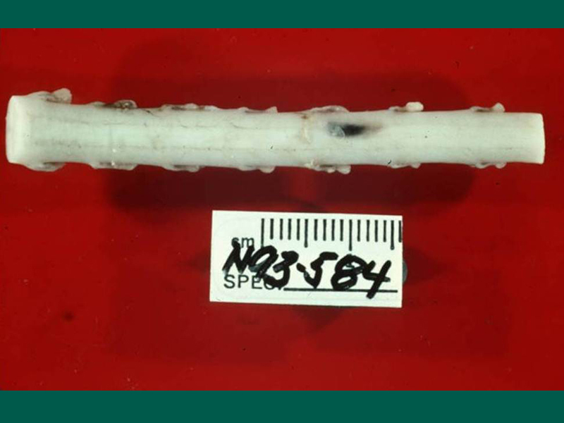
Spinal cord lesion due to Cuterebra maggot migrating through the spinal cord of a cat
Disease
- Clinical signs are usually not apparent in the normal rodent or lagomorph host.
- In cats and dogs, the presence of clinical signs depends largely on where the larvae have migrated (e.g. nares, upper respiratory tract, skin, eye, brain).
- There are three forms of disease:
- Furuncular myiasis: subcutaneous cysts with mature third-instar bots in the skin
- Neurologic and other disease manifestations from the migration of young bots through deeper tissues of the body
- Respiratory distress or upper respiratory disease from migration of young bots through the trachea and diaphragm
- The most common presentation is a subcutaneous cyst which has a 2 to 4 mm opening with well defined margins and serous discharge.
- Most cysts containing bots are found around the face or neck
- Rarely, larvae may enter the anterior chamber or globe of the eye causing chemosis, blepharospasm, serous ocular discharge, and exudative uveitis with blindness
- Typically, there are no signs of disease or distress in cats or dogs due to larvae localized subcutaneously
- Most cysts containing bots are found around the face or neck
- Neurologic disease, which is reported more often in cats than dogs, occurs when larvae migrate through the brain
- Clinical signs vary from acute onset of status epilepticus with no recovery, to multiple signs (head tilt, unilateral or bilateral central blindness, head pressing, cognitive dysfunction, continuous vocalization, proprioceptive deficits, circling) indicating multifocal CNS lesions, to only severely depressed mentation. After an acute onset, signs are usually rapidly progressive and ultimately fatal; more rarely, some cases may progress for weeks or months.
- Some cats, after an acute onset of cerebral signs, recover and are left with residual signs of abnormal behavior, e.g., constant pacing or circling, abnormal mentation, or seizures. Occasionally spontaneous recovery appears to be complete.
- Neurologic signs typically develop 1 to 2 weeks after respiratory signs have been noted, although respiratory signs may occur as long as 4 to 10 weeks before the onset of neurologic disease.
- In cats with respiratory disease, some present with a history of sneezing and nasal discharge, unilateral facial swellings especially over the nose, extreme respiratory dyspnea sometimes with a bloody nasal discharge, and soft palate and pharyngeal swelling.
- Unlike cutaneous lesions, respiratory signs more often precede the development of neurologic signs.
Prevalence
The prevalence of infection in rodent and rabbit populations may be as high as 30% to 70% in some areas but is much lower in dogs and cats in the same area.
Host Associations and Transmission Between Hosts
Cats and dogs are accidental hosts infected when a first-stage maggot enters through the mouth, eyes, anus, vulva, or nares. Infection does not occur due to skin penetration.
Although mature third-stage instars in dogs and cats are fully viable and able to pupate after emerging from the cyst, these bots are usually incapable of maturing into adult flies. Thus, dogs and cats do not appear to play any role as reservoir hosts.
Once a bot is inside the body of its host, there is no host-to-host transmission. Infection only occurs via the entrance of a newly hatched larva into a susceptible host.
Prepatent Period and Environmental Factors
- It takes 3 to 6 weeks from infection for the newly hatched larva to develop into a fully formed third-stage larva in dogs and cats
- In warmer climates, adult flies can emerge from soil and lay eggs throughout the year. Thus, there is no seasonality associated with observed cases.
- In areas with cold winters, cases tend to occur in the summer and early fall due to the fact that the flies lay eggs only at one time of the year.
- Cases may occur in winter months when eggs are maintained within some protected location.
Site of Infection and Pathogenesis
- Subcutaneous tissue, nervous tissue (brain or spinal cord), or respiratory tissues are the most common sites where larvae are found.
- Infections of the eye are also possible.
- In the skin, the cyst around the larva contains a thin layer of nectrotic tissue on which the bot feeds.
- Migration of young bots through the esophagus, trachea and diaphragm causes mechanical damage and inflammation in the upper respiratory tract.
- Cuterebra infection seems to be a cause of feline ischemic encephalopathy, and it has been postulated that the vascular spasm associated with feline ischemic syndrome may be due to some toxin produced by the circulating fly larva.
Diagnosis
- A Cuterebra bot in a subcutaneous cyst is generally distinctive and diagnostic as these are highly host- and site-specific parasites.
- Larvae causing neurologic or respiratory signs represent more of a diagnostic challenge.
- Suspicion of infection should be raised by acute upper respiratory disease (especially with unilateral signs of nasal discharge or nasal/facial swelling), or acute onset of neurologic disease possibly preceeded by upper respiratory signs 1 to 2 weeks previously.
- These signs may be accompanied by a peripheral eosinophilia.
- CAT scans of an affected dog or cat may reveal a mottled appearance to the brain consistent with encephalitis, but no conclusive evidence of infection
- Magnetic resonance imaging may reveal larvae or their migratory tracks in the brain or spinal cord
- In cases presenting with upper respiratory disease, examination of the pharynx, larynx, and nasal passages under general anesthesia may reveal a larva.
- An enzyme-linked immunosorbent assay has recently been developed, using crude somatic antigens from third-stage instars that can detect parasite-specific IgG in infected cats; however, because the neurologic disease is often acute, IgG may not yet be present in sufficient quantities for detection ([email protected]).
- During necropsy of cats with neurologic disease, larvae may be observed intracranially or within the spinal cord.
Treatment
- Subcutaneous bots
- Surgical extraction of the bot(s)
- Mature bots within a cyst can be removed by expanding the cyst opening and carefully extracting with forceps; care should be taken to avoid crushing the bot during removal.
- Smaller, less developed larvae within the skin require removal by careful dissection.
- Surgical extraction of the bot(s)
- Larvae within the soft tissues of the mouth, nasal sinus, larynx, or eye require surgical extraction.
- Successful removal of larvae that are located intracranially or within the spinal cord has not been reported.
- Ivermectin has been shown to be effective against migrating larvae of Cuterebra at 0.1 – 0.3 mg/kg. Although prognosis is guarded in cats with neurologic signs, treatment of cats with Cuterebra-associated respiratory disease with a combination of ivermectin and corticosteroids may be of clinical value.
Control and Prevention
Cats and dogs with access to outdoor areas where infected rodents and rabbits are present are at risk.
No products are labeled for control of Cuterebra spp. However, topical insecticides, such as fipronil and imidacloprid, may provide some protection from infestation. Similarly, administering systemic macrocyclic lactones may kill larvae during migration.
Public Health Considerations
- The larvae in the cat or dog does not pose a health risk to humans.
- There have been reports of the recovery of larvae of Cuterebra from humans
- Humans, like cats and dogs, are infected by larvae entering an orifice and notvia skin penetration as occurs with Dermotobia hominis and Hypoderma species.
Selected References
- Cansi ER. 2011. Record of Myiasis by Cuterebra apicalis in Domestic Dog in the Central Region of Brazil (Article in Portuguese). Acta Scientae Vet 39(2): e969, 3 pages
- Crumley WR, Rankin AJ, Dryden MW. 2011. Ophthalmomyiasis externa in a puppy due to Cuterebra infestation. J Am Anim Hosp Assoc. 47(6):e150-155.
- Glass EN, Cornetta AM, deLahunta A, Center SA, Kent M. 1998. Clinical and clinicopathologic features in 11 cats with Cuterebra larvae myiasis of the central nervous system. J Vet Int Med 12:365-368.
- Tieber LM, Axlund TW, Simpson ST, Hathcock JT. 2006. Survival of a suspected case of central nervous system cuterebrosis in a dog: clinical and magnetic resonance imaging findings. J Am Anim Hosp Assoc. 42:238-242.
- Williams KJ, Summers BA, de Lahunta A. 1998. Cerebrospinal cuterebriasis in cats and its association with feline ischemic encephalopathy. Vet Pathol 35:330-343.